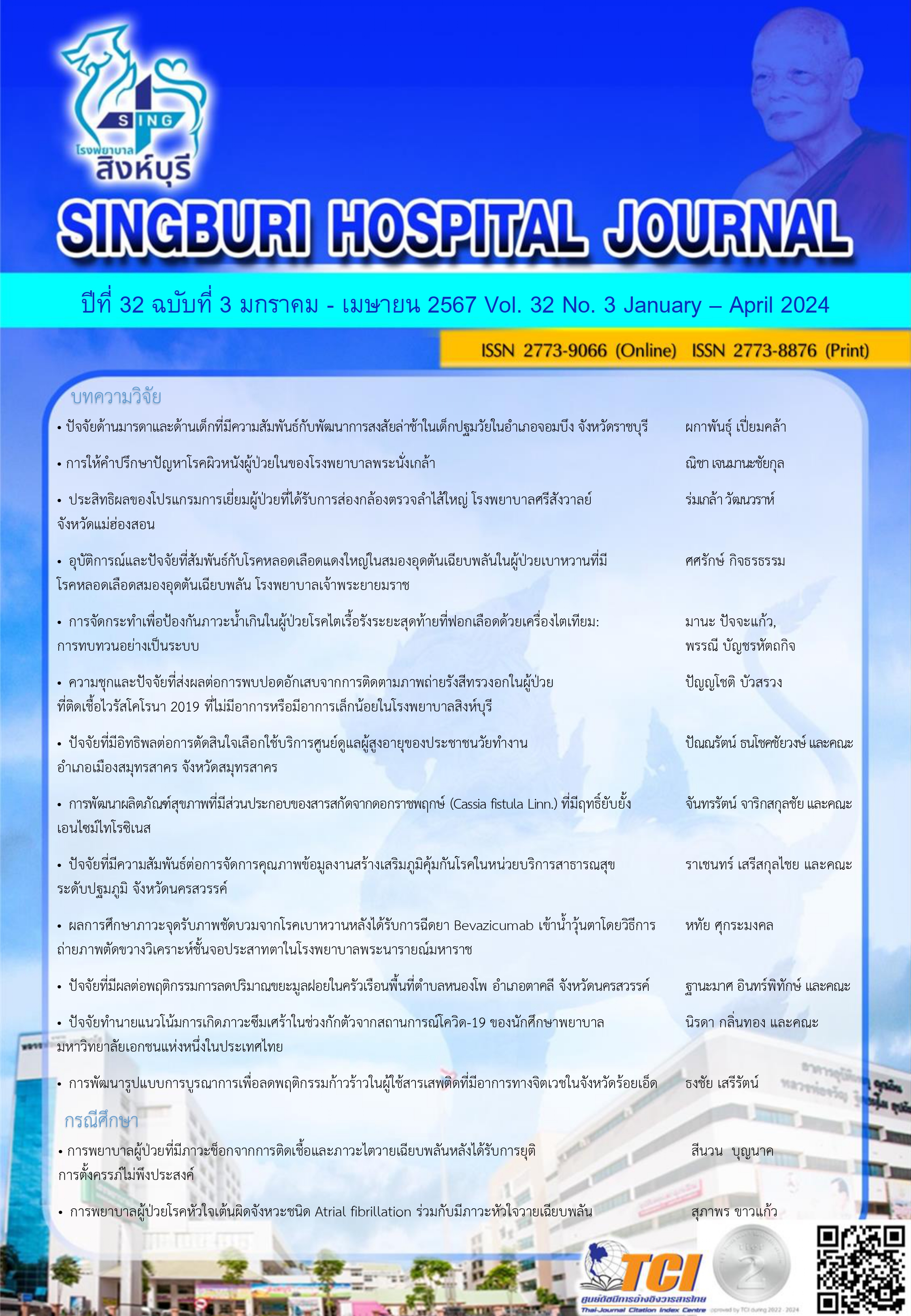
ความชุกและปัจจัยที่ส่งผลต่อการพบปอดอักเสบจากการติดตามภาพถ่ายรังสีทรวงอก ในผู้ป่วยที่ติดเชื้อไวรัสโคโรนา 2019 ที่ไม่มีอาการหรือมีอาการเล็กน้อยในโรงพยาบาลสิงห์บุรี
คำสำคัญ:
ปอดอักเสบ, ไวรัสโคโรนา 2019, ภาพถ่ายรังสีทรวงอก, การติดตามบทคัดย่อ
การตรวจด้วยภาพรังสีทรวงอกเป็นวิธีการตรวจเบื้องต้นสำหรับการวินิจฉัยในกลุ่มผู้ป่วยที่ติดเชื้อไวรัสโคโรนา 2019 ทั้งนี้ยังไม่มีข้อมูลบทบาทของตรวจติดตามด้วยภาพรังสีทรวงอกของในกลุ่มที่ไม่มีอาการหรือมีอาการเล็กน้อย การศึกษาครั้งนี้มีวัตถุประสงค์เพื่อศึกษาความชุกและปัจจัยที่ส่งผลต่อการพบปอดอักเสบจากการติดตามภาพรังสีทรวงอกครั้งที่ 2 ในกลุ่มผู้ป่วยที่ติดเชื้อไวรัสโคโรนา 2019 ที่ไม่มีอาการหรือมีอาการเล็กน้อย การศึกษาครั้งนี้เป็นการทบทวนเวชระเบียนเชิงพรรณนาในผู้ป่วยที่ติดเชื้อไวรัสโคโรนา 2019 รายใหม่ที่ไม่มีอาการหรือมีอาการเล็กน้อย จำนวนทั้งสิ้น 717 ราย ที่เข้ารับการรักษาที่โรงพยาบาลสิงห์บุรีหรือโรงพยาบาลสนาม ตั้งแต่วันที่ 1 กันยายน พ.ศ. 2564 ถึงวันที่ 28 กุมภาพันธ์ พ.ศ. 2565 วิเคราะห์ข้อมูลด้วยสถิติถดถอยพหุโลจิสติก (multiple logistic regression analysis)
ผลการศึกษาพบว่าค่ามัธยฐานของอายุอยู่ที่ 37 ปี เป็นเพศหญิง 420 ราย (58%) ได้รับวัคซีนไวรัสโคโรนา-2019 จำนวน 522 ราย (73%) มีโรคประจำตัว 309 ราย (43%) เป็นโรคอ้วน 177 ราย (24%) มีความชุกของผู้ป่วยที่มีปอดอักเสบรายใหม่จากการติดตามภาพรังสีทรวงอกครั้งที่ 2 อยู่ที่ 108 ราย (15%) ปัจจัยที่ส่งผลต่อการพบปอดอักเสบจากการติดตามภาพรังสีทรวงอกครั้งที่ 2 ได้แก่ อายุ ดัชนีมวลกาย การได้รับวัคซีนป้องกันไวรัสโคโรนา 2019 มีโรคประจำตัวไตวายเรื้อรังตั้งแต่ระยะที่ 3 มีอาการท้องร่วง ระดับ CRP AST และ ALT สูงผิดปกติ
Downloads
เอกสารอ้างอิง
World Health Organization. Weekly epidemiological update on COVID-19 - 1 March 2023 [Internet]. 2023 [updated 2023 Mar 1; cited 2023 Mar 3]. Available from: https://www.who.int/publications/m/item/weekly-epidemiological-update-on-covid-19---1-march-2023.
Department of Disease Control, Ministry of Public Health (TH), Coronavirus disease (COVID-19): Thailand situation 2023 [Internet]. 2023 [updated 2023 Mar 1; cited 2023 Mar 3]. Available form: https://ddc.moph.go.th/viralpneumonia.
Zhang JJ, Dong X, Liu GH, Gao DY. Risk and protective factors for COVID-19 morbidity, severity, and mortality. Clin Rev Allergy Immunol. 2023;64(1):90-107.
กรมการแพทย์ กระทรวงสาธารณสุข. แนวทางเวชปฏิบัติการวินิจฉัย ดูแลรักษา และป้องกันการติดเชื้อในโรงพยาบาล กรณีโรคติดเชื้อไวรัสโคโรนา 2019 (COVID-19) ฉบับปรับปรุงวันที่ 18 เมษายน พ.ศ. 2566 สำหรับแพทย์และบุคลากรสาธารณสุข [อินเตอร์เน็ต]. 2566 [เข้าถึงเมื่อ 28 เมษายน 2566]; เข้าถึงได้จาก: https://covid19.dms.go.th/backend///Content//Content_File/Covid_Health/Attach/25650301194159PM_CPG_COVID-19_v.20.4_N_20220301.pdf
Prutipinyo C. COVID-19 field hospital: alternative state quarantine hospital and hospitel. Public Health Policy and Laws Journal. 2021;7:195-213.
Department of Medicine Services, Ministry of Public Health (TH), Guidelines on triage individuals with confirmed COVID-19 in Bangkok [Internet]. 2023 [updated 2021 Nov 30; cited 2023 Mar 3]. Available form: https://covid19.dms.go.th/Content/Select_Landding_page?contentId=120.
National Institutes of Health. COVID-19 Treatment Guidelines Panel. Coronavirus Disease 2019 (COVID-19) Treatment Guidelines. 2021 [cited2021 Nov 30] Available form: https://www.covid19treatmentguidelines.nih.gov/
World Health Organization. Clinical management of COVID-19 (WHO/2019-nCoV/clinical/2020.5). 2020. [Cited 2021 Nov 30] Available form: https://www.who.int/publications/i/ item/clinical-management-of-covid-19
Liqa AR, Eyhab E, Musaab K, et al. Chest x-ray findings and temporal lung changes in patients with COVID-19 pneumonia. BMC Pulm Med. 2022;20:245.
Rahab Y, Walaa G. Chest X-ray findings monitoring COVID-19 disease course and severity. Egypt J Radiol Nucl Med. 2021; 52:193.
Kuo BJ, Lai YK, Tan MLM, et al. Utility of screening chest radiographs in patients with asymptomatic or minimally symptomatic COVID-19 in Singapore. Radiology. 2021; 298(3):E131-40.
Trimankha P, Lakkana J, Rungsin R, et al. Utility of screening chest radiographs in patients with asymptomatic and mildly symptomatic COVID-19 at a field hospital in Samut Sakhon, Thailand. ASEAN J Radiol. 2021;22(2):5-20.
Mostafa MK, Hassan AS, Abdullah SA, et al. Early prediction keys for COVID-19 cases progression: A meta-analysis. J Infect Public Health. 2021; 14(5):561-9.
Özger HS, Aysert Yıldız P, Gaygısız Ü, et al. The factors predicting pneumonia in COVID-19 patients: preliminary results from a university hospital in Turkey. Turk J Med Sci. 2020;50:1810–6.
Cordero-Franco HF, De La Garza-Salinas LH, Gomez-Garcia S, et al. Risk factors for SARS-CoV-2 infection, pneumonia, intubation, and death in Northeast Mexico. Front Public Health. 2021;9:645739.
ฐาปกรณ์ จิตตนูนท์. การทำนายความเสี่ยงของการเกิดโรคปอดอักเสบในผู้ป่วยโควิด-19 ที่โรงพยาบาลบางปะอิน จังหวัดพระนครศรีอยุธยา. วารสารสมาคมเวชศาสตร์ป้องกันแห่งประเทศไทย. 2566;13(1):30-45.
Chenchula S, Vidyasagar K, Pathan S. et al. Global prevalence and effect of comorbidities and smoking status on severity and mortality of COVID-19 in association with age and gender: a systematic review, meta-analysis and meta-regression. Sci Rep. 2023;13:6415.
Rahmani K, Shavaleh R, Forouhi M, et al. The effectiveness of COVID-19 vaccines in reducing the incidence, hospitalization, and mortality from COVID-19: A systematic review and meta-analysis. Front Public Health. 2022; 10:873596.
Jdiaa SS, Mansour R, El Alayli A, et al. COVID-19 and chronic kidney disease: an updated overview of reviews. J Nephrol. 2022;35:69-85.
Ghimire S, Sharma S, Patel A, et al. Diarrhea Is Associated with Increased Severity of Disease in COVID-19: Systemic Review and Metaanalysis. SN Compr Clin Med. 2021;3:28-35.
Ali N. Elevated level of C-reactive protein may be an early marker to predict risk for severity of COVID-19. J Med Virol. 2020;92(11):2409–11.
Wang L. C-Reactive protein levels in the early stage of COVID-19. Med Mal Infect. 2020;50(4):332–4.
Goyal D, Inada-Kim M, Mansab F, et al. improving the early identification of COVID-19 pneumonia: a narrative review. BMJ Open Respir Res. 2021;8(1):e000911.
Krishnan A, Prichett L, Tao X, et al. Abnormal liver chemistries as a predictor of COVID-19 severity and clinical outcomes in hospitalized patients. World J Gastroenterol. 2022;28(5):570-87.
Lee JY, Nam BH, Kim M, Hwang J, Kim JY, et al. A risk scoring system to predict progression to severe pneumonia in patients with Covid-19. Sci Rep. 2022;12(1):5390.
Medetalibeyoglu A, Catma Y, Senkal N, Ormeci A, Cavus B, et al. The effect of liver test abnormalities on the prognosis of COVID-19. Ann Hepatol. 2020;19(6):614-21.
ดาวน์โหลด
เผยแพร่แล้ว
รูปแบบการอ้างอิง
ฉบับ
ประเภทบทความ
สัญญาอนุญาต
ลิขสิทธิ์ (c) 2024 โรงพยาบาลสิงห์บุรี

อนุญาตภายใต้เงื่อนไข Creative Commons Attribution-NonCommercial-NoDerivatives 4.0 International License.
บทความที่ได้รับการตีพิมพ์เป็นลิขสิทธิ์ของโรงพยาบาลสิงห์บุรี
ข้อความที่ปรากฏในบทความแต่ละเรื่องในวารสารวิชาการเล่มนี้เป็นความคิดเห็นส่วนตัวของผู้เขียนแต่ละท่านไม่เกี่ยวข้องกับโรงพยาบาลสิงห์บุรี และบุคคลากรท่านอื่นๆในโรงพยาบาลฯ แต่อย่างใด ความรับผิดชอบองค์ประกอบทั้งหมดของบทความแต่ละเรื่องเป็นของผู้เขียนแต่ละท่าน หากมีความผิดพลาดใดๆ ผู้เขียนแต่ละท่านจะรับผิดชอบบทความของตนเองแต่ผู้เดียว


