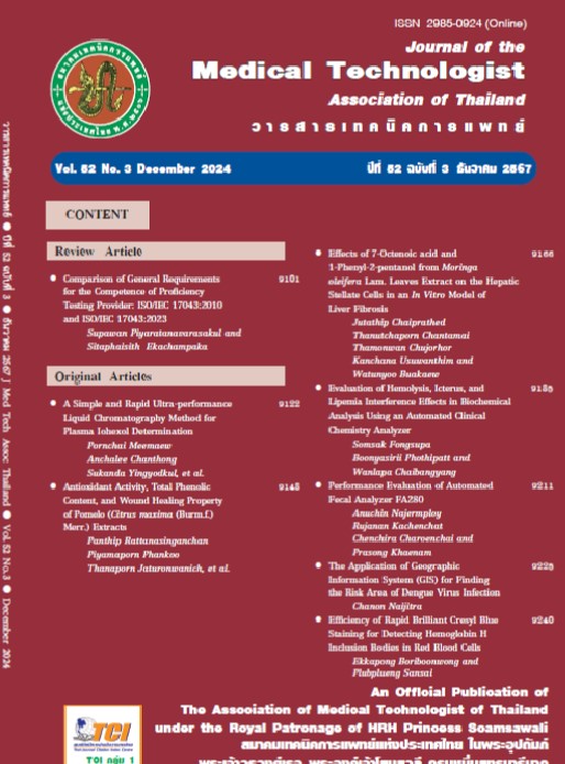
การประเมินผลกระทบจากการแตกของเม็ดเลือดแดง ความเหลือง และความขุ่น ต่อการวิเคราะห์สารชีวเคมี ด้วยเครื่องวิเคราะห์อัตโนมัติทางเคมีคลินิก
คำสำคัญ:
การรบกวน, การแตกของเม็ดเลือดแดง, ความเหลือง, ความขุ่น, HIL indexบทคัดย่อ
สิ่งส่งตรวจประเภทซีรัมหรือพลาสมาที่มีการแตกของเม็ดเลือดแดง (hemolysis, H) มีความเหลืองจากบิลิรูบิน (icterus, I) หรือมีความขุ่น (lipemia, L) เป็นปัญหาในขั้นตอนก่อนการวิเคราะห์ซึ่งส่งผลกระทบต่อค่าการตรวจวัด ที่พบเป็นประจำในห้องปฏิบัติการเคมีคลินิก ปัจจุบันการประเมิน HIL index ในสิ่งส่งตรวจด้วยเครื่องวิเคราะห์อัตโนมัติทางเคมีคลินิกโดยวิธีวัดการดูดกลืนแสง เป็นวิธีที่มีความน่าเชื่อถือและนิยมใช้กันแพร่หลาย การศึกษานี้จึงมีวัตถุประสงค์เพื่อประเมินผลการรบกวนจาก HIL ต่อการวิเคราะห์สารชีวเคมีในเลือดด้วยเครื่องวิเคราะห์อัตโนมัติทางเคมีคลินิก Beckman Coulter AU5800 โดยใช้ข้อมูลผลการประเมิน HIL index เชิงกึ่งปริมาณและเชิงปริมาณ โดยเก็บข้อมูลจากผลการวิเคราะห์ระดับสารชีวเคมีของตัวอย่างผู้ป่วยระหว่างเดือนมิถุนายน-ธันวาคม พ.ศ. 2566 เพื่อเปรียบเทียบความแตกต่างของระดับสารชีวเคมีที่เปลี่ยนแปลงไปตามระดับของ HIL index และศึกษาความสัมพันธ์ระหว่างระดับสารชีวเคมีกับ HIL index เชิงปริมาณเพื่อทำนายระดับ HIL index ที่มีการรบกวนการวิเคราะห์โดยใช้ตัวแบบถดถอยเชิงเส้น จากผลการวิจัยพบว่าจากข้อมูล HIL index สามารถประเมินผลการรบกวนได้สูงสุดที่ระดับ (2+) โดยพบการรบกวนเชิงบวกจากการแตกของเม็ดเลือดแดง (hemolysis) อย่างมีนัยสำคัญทางคลินิกในการวิเคราะห์ lactate dehydrogenase (LDH) และ aspartate aminotransferase (AST) ที่ดัชนีการแตกของเม็ดเลือดแดง (HI) ระดับ (1+) และการรบกวนเชิงลบในการวิเคราะห์ total bilirubin (TB) ที่ HI ระดับ (2+) นอกจากนี้พบการรบกวนเชิงบวกจากความขุ่น (lipemia) อย่างมีนัยสำคัญทางคลินิกในการวิเคราะห์ AST ที่ดัชนีความขุ่น (LI) ระดับ (2+) ไม่พบการรบกวนอย่างมีนัยสำคัญทางคลินิกในการวิเคราะห์ alanine aminotransferase (ALT), direct bilirubin (DB), blood urea nitrogen (BUN), และ magnesium จนถึงระดับ HI (2+) การวิเคราะห์ cholesterol จนถึงดัชนีความเหลืองจากบิลิรูบิน (II) ระดับ (1+) และการวิเคราะห์ ALT จนถึงระดับ LI (2+) ทั้งนี้ผลการทำนายระดับที่พบการรบกวนจากการแตกของเม็ดเลือดแดง (hemolysis) โดยใช้ตัวแบบถดถอยเชิงเส้นของการวิเคราะห์ LDH ช่วยระบุ cutoff ที่มีการรบกวนตั้งแต่ระดับ HI (0) ได้ ดังนั้น ห้องปฏิบัติการควรประเมินและกำหนดระดับค่า cutoff ที่เหมาะสมของ HIL index เพื่อประโยชน์ในการตัดสินใจดำเนินการกับสิ่งส่งตรวจและผลการวิเคราะห์ เพื่อให้ผู้ป่วยได้รับความปลอดภัยสูงสุดจากผลตรวจทางห้องปฏิบัติการ
เอกสารอ้างอิง
Chawla R, Goswami B, Tayal D, Mallika V. Identification of the types of preanalytical errors in the clinical chemistry laboratory: 1-year study at G.B. Pant Hospital. Lab Med 2010; 41: 89-92.
Ryder K, Glick M and Glick S. Incidence and amount of turbidity, hemolysis, and icterus in serum from outpatients. Lab Med 1991; 22: 415-8.
Kroll MH, McCudden CR. Endogenous interferences in clinical laboratory tests: icteric, lipemic and turbid samples. Berlin:De Gruyter; 2012.
Marques-Garcia F. Methods for hemolysis interference study in laboratory medicine - a critical review. eJIFCC 2020; 31: 85-97.
Farrell CJ, Carter AC. Serum indices: managing assay interference. Ann Clin Biochem. 2016; 53: 527-38.
Dimeski G, Mollee P, Carter A. Effects of hyperlipidemia on plasma sodium, potassium, and chloride measurements by an indirect ion-selective electrode measuring system. Clin Chem 2006; 52: 155-6.
Glick MR, Ryder KW, Glick SJ, Woods JR. Unreliable visual estimation of the and amount of turbidity, hemolysis, and icterus in serum from patients. Clin Chem 1989; 35: 837-9.
Lippi G, Cadamuro J. Visual assessment of sample quality: quo usque tandem?. Clin Chem Lab Med 2018; 56: 513-5.
Simundic AM, Nikolac N, Ivankovic V, et al. Comparison of visual vs. Automated detection of lipemic, icteric and hemolyzed specimens: can we rely on a human eye?. Clin Chem Lab Med 2009; 47: 1361-5.
Getahun T, Alemu A, Mulugeta F, et al. Evaluation of visual serum indices measurements and potential false result risks in routine clinical chemistry tests in Addis Ababa, Ethiopia. eJIFCC 2019; 30: 276-87.
von Meyer A, Cadamuro J, Lippi G, Simundic AM. Call for more transparency in manufacturers declarations on serum indices: On behalf of the Working Group for Preanalytical Phase (WG-PRE), European Federation of Clinical Chemistry and Laboratory Medicine (EFLM). Clin Chim Acta 2018; 484: 328-32.
Clinical and Laboratory Standards Institute (CLSI). Hemolysis, icterus, and lipemia/ turbidity indices as indicators of interference in clinical laboratory analysis; approved guideline. C56-A. Wayne, PA: Clinical and Laboratory Standards Institute, 2012.
Lim YK, Young JC. Proposal of modified HIL-indices for determining hemolysis, icterus and lipemia interference on the Beckman Coulter AU5800 automated platform. Lab Med Online 2017; 7: 66-72.
Aslan B, Stemp J, Catomeris P, Allen L, Kerekes R, Gun-Munro J. Clinical chemistry sample interferences reporting patterns in Ontario laboratories (abstract). Clin Chem 2012; 58: A23-4.
Akbas N, Eppert B, Miller J, Schulten C, Wallace C, Turner T. Effect of hemolysis, icterus and lipemia on chemistry tests and association between the amount of interfering substances and LIH indices. Medpace Reference Laboratories, Cincinnati, OH; 2018 [cited 2024 Feb 5]. Available from: https://www.medpace.com/wp-content/uploads/2018/08/Whitepaper-LIH.pdf
Beckman Coulter Inc. LIH setting sheet (technical document). Brea (CA): Beckman Coulter Inc, 2022 [cited 2024 Feb 5]. Available from: https://www.beckmancoulter.com/download/file/phx-BSOSR6X16604-EN_US/BSOSR6X16604?type=pdf
Box GEP, Cox DR. An analysis of transformations. J R Stat Soc 1964; B26: 211-52.
Horn PS, Feng L, Li Y, Pesce AJ. Effect of outliers and nonhealthy individuals on reference interval estimation. Clin Chem 2001; 47: 2137-45.
Jenkins DG, Quintana-Ascencio PF. A solution to minimum sample size for regressions. PLoS One 2020; 15: e0229345.
Aarsand AK, Fernandez-Calle P, Webster C, et al. The EFLM Biological Variation Database [cited 2024 Jan 14]. Available from: https://biologicalvariation.eu/
Ricós C, Alvarez V, Cava F, et al. Current databases on biological variation: pros, cons and progress. Scand J Clin Lab Invest. 1999; 59: 491-500 [cited 2024 Feb 5]. Updated database available from: https://www.westgard.com/clia-a-quality/quality-requirements/238-biodatabase1.html
Tian G, Wu Y, Jin X, et al. The incidence rate and influence factors of hemolysis, lipemia, icterus in fasting serum biochemistry specimens. PLoS One 2022; 17: e0262748.
Perović A, Dolčić M. Influence of hemolysis on clinical chemistry parameters determined with Beckman Coulter tests - detection of clinically significant interference. Scand J Clin Lab Invest 2019; 79: 154-9.
Koseoglu M, Hur A, Atay A, Cuhadar S. Effects of hemolysis interferences on routine biochemistry parameters. Biochem Med 2011; 21: 79-85.
Lippi G, Salvagno GL, Montagnana M, Brocco G, Guidi GC. Influence of hemolysis on routine clinical chemistry testing. Clin Chem Lab Med 2006; 44:311-6.
Ho CKM, Chen C, Setoh JWS, Yap WWT, Hawkins RCW. Optimization of hemolysis, icterus and lipemia interference thresholds for 35 clinical chemistry assays.Pract Lab Med 2021; 25: e00232.
Shull B, Lees H, Li P. Mechanism of interference by hemoglobin in the determination of total bilirubin. I. Method of Malloy-Evelyn. Clin Chem 1980; 26: 22-5.
Dawson R. How significant is a boxplot outlier?. J Stat Educ 2011; 19.
Fernández-Prendes C, Castro-Castro MJ, Jiménez-Añón L, Morales-Indiano C, Martínez-Bujidos M. Discrepancies in lipemia interference between endogenous lipemic samples and Smoflipid®-supplemented samples. eJIFCC 2023; 34:27-41.
ดาวน์โหลด
เผยแพร่แล้ว
รูปแบบการอ้างอิง
ฉบับ
ประเภทบทความ
สัญญาอนุญาต
ลิขสิทธิ์ (c) 2024 วารสารเทคนิคการแพทย์

อนุญาตภายใต้เงื่อนไข Creative Commons Attribution-NonCommercial-NoDerivatives 4.0 International License.


