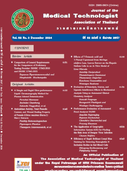
ฤทธิ์การต้านพังผืดในตับของสารออกฤทธิ์ 7-Octenoic acid และ 1-Phenyl-2-pentanol จากใบมะรุม
คำสำคัญ:
พังผืดในตับ, มะรุม, กรดออกทาโนอิก, 1-ฟีนิล-2-เพนทานอลบทคัดย่อ
ภาวะพังผืดในตับ (liver fibrosis) คือภาวะที่มีการสะสมของ extracellular matrix protein ในตับที่มากเกินไป ซึ่งสาเหตุที่พบบ่อย ได้แก่ ภาวะไขมันพอกตับและโรคไวรัสตับอักเสบซี หากไม่ได้รับการรักษาที่ถูกต้อง จะทำให้เพิ่มโอกาสเสี่ยงต่อการเกิดภาวะตับแข็งและมะเร็งตับ ปัจจุบันภาวะพังผืดในตับยังไม่มียารักษาที่เป็นมาตรฐาน มีการศึกษาพบว่าสารสกัดจากมะรุมมีฤทธิ์ในการต้านการอักเสบในตับได้ คณะผู้วิจัยจึงสนใจที่จะศึกษากลไกการต้านการเกิดพังผืดในตับของสารออกฤทธิ์ที่แยกได้จากใบมะรุม 2 ชนิด ได้แก่ 7-octenoic acid และ 1-phenyl-2-pentanol ในเซลล์ตับชนิด hepatic stellate cells (HSCs) โดยวัดระดับไซโตไคน์ด้วยวิธี sandwich ELISA และวิเคราะห์การแสดงออกของโปรตีนภายในเซลล์ HSCs ด้วยเทคนิคโปรตีโอมิกส์ ผลการศึกษาพบว่าเซลล์ HSCs ที่ได้รับ 7-octenoic acid และ 1-phenyl-2-pentanol มีการเปลี่ยนแปลงระดับการแสดงออกของโปรตีนที่เพิ่มขึ้นและลดลงจำนวน 440 และ 98 ชนิด ตามลำดับ อย่างมีนัยสำคัญทางสถิติ (p < 0.05) เมื่อเปรียบเทียบกับกลุ่มควบคุม อีกทั้งพบการลดลงของการแสดงออกของโปรตีน casein kinase II subunit alpha (CSNK2A1), guanine nucleotide-binding protein G(I)/G(S)/G(T) subunit beta-2 (GNB2) และ low-density lipoprotein receptor-related protein 5 (LRP5) ซึ่งเป็นโปรตีนที่มีความเกี่ยวข้องกับวิถีสัญญาณ NF-κB pathway, PI3K/Akt signaling pathway และ mTOR signaling pathway นอกจากนี้ 7-octenoic acid และ 1-phenyl-2-pentanol ยังสามารถลดการหลั่งของ TNF-α ได้อย่างมีนัยสำคัญทางสถิติ (p < 0.05) โดยผลการศึกษานี้พบว่า สารทั้งสองชนิดมีคุณสมบัติลดการกระตุ้นสัญญาณ TGF-β1 ต่อเซลล์ตับชนิด HSCs ได้ ซึ่งมีความน่าสนใจสำหรับการศึกษาต่อเพื่อนำไปใช้ในการพัฒนาเป็นยาต้านการเกิดพังผืดในตับในอนาคต
เอกสารอ้างอิง
Bataller R, Brenner DA. Liver fibrosis. J Clin Invest 2005; 115: 209-18.
Menggensilimu, Yuan H, Zhao C, et al. Anti-liver fibrosis effect of total flavonoids from Scabiosa comosa Fisch. ex Roem. et Schult. on liver fibrosis in rat models and its proteomics analysis. Ann Palliat Med 2020; 9: 272-85.
Sarin SK, Kumar M, Eslam M, et al. Liver diseases in the Asia-Pacific region: a Lancet Gastroenterology & Hepatology Commission. Lancet Gastroenterol Hepatol 2020; 5: 167-228.
Poynard T, Lebray P, Ingiliz P, et al. Prevalence of liver fibrosis and risk factors in a general population using non-invasive biomarkers (FibroTest). BMC Gastroenterol 2010; 10: 1-13.
Khanam A, Saleeb PG, Kottilil S. Pathophysiology and treatment options for hepatic fibrosis: can it be completely cured? Cells 2021; 10: 1097.
Yang YM, Seki E. TNFoc in liver fibrosis. Curr Pathobiol Rep 2015; 3: 253-61.
Zelova H, Hosek J. TNF-alpha signalling and inflammation: interactions between old acquaintances. Inflamm Res 2013; 62: 641-51.
Tan Z, Sun H, Xue T, et al. Liver fibrosis: therapeutic targets and advances in drug therapy. Front Cell Dev Biol 2021; 9: 730176.
Camellia Seed Oil and Other Plant Oil Product Research and Development Center. (MORINGA) Chiang Rai, Thailand: [Available from: https://www.camelliaoilcenter.com/%E0%B8%A1%E0%B8%B0%E0%B8%A3%E0%B8%B8%E0%B8%A1-moringa/.
Kou X, Li B, Olayanju JB, Drake JM, Chen N. Nutraceutical or pharmacological potential of Moringa oleifera Lam. Nutrients 2018; 10: 343.
Wisitpongpun P, Suphrom N, Potup P, et al. In vitro bioassay-guided identification of anticancer properties from Moringa oleifera Lam. leaf against the MDAMB-231 cell line. Pharmaceuticals 2020;13: 464.
Yu J, Hu Y, Gao Y, et al. Kindlin-2 regulates hepatic stellate cells activation and liver fibrogenesis. Cell Death Discovery 2018; 4: 93.
Xu Z, Li T, Li M, et al. eRF3b-37 inhibits the TGF-β 1-induced activation of hepatic stellate cells by regulating cell proliferation, G0/G1 arrest, apoptosis and migration. Int J Mol Med 2018; 42: 3602-12.
Wei S, Wang Q, Zhou H, et al. miR-455- 3p alleviates hepatic stellate cell activation and liver fibrosis by suppressing HSF1 expression. Mol Ther Nucleic Acids 2019; 16: 758-69.
Wang Y, Wu C, Zhou J, Fang H, Wang J. Overexpression of estrogen receptor β inhibits cellular functions of human hepatic stellate cells and promotes the anti-fibrosis effect of calycosin via inhibiting STAT3 phosphorylation. BMC Pharmacol Toxicol 2022; 23: 77.
Kang H, Seo E, Oh YS, Jun H-S. TGF-β activates NLRP3 inflammasome by an autocrine production of TGF-β in LX-2 human hepatic stellate cells. Mol Cell Biochem 2022; 477: 1329-38.
Buakaew W, Krobthong S, Yingchutrakul Y, et al. Investigating the antifibrotic effects of β-Citronellol on a TGF-β 1-stimulated LX-2 hepatic stellate cell Model. Biomolecules 2024; 14: 800.
Szklarczyk D, Franceschini A, Wyder S, et al. STRING v10: protein–protein interaction networks, integrated over the tree of life. Nucleic Acids Res 2015; 43: 447-52.
Chen EY, Tan CM, Kou Y, et al. Enrichr: interactive and collaborative HTML5 gene list enrichment analysis tool. BMC Bioinformatics 2013; 14: 128.
Kuleshov MV, Jones MR, Rouillard AD, et al. Enrichr: a comprehensive gene set enrichment analysis web server 2016 update. Nucleic Acids Res 2016; 44: 90-7.
Xie Z, Bailey A, Kuleshov MV, et al. Gene set knowledge discovery with Enrichr. Current Protocols 2021; 1: 90.
Kanehisa M, Goto S. KEGG: Kyoto encyclopedia of genes and genomes. Nucleic Acids Res 2000; 28: 27-30.
Kanehisa M. Toward understanding the origin and evolution of cellular organisms. Protein Science 2019; 28: 1947-51.
Kanehisa M, Furumichi M, Sato Y, Kawashima M, Ishiguro-Watanabe M. KEGG for taxonomy-based analysis of pathways and genomes. Nucleic Acids Res 2023; 51: 587-92.
Jodynis-Liebert J, Kujawska M. Biphasic dose-response induced by phytochemicals: experimental evidence. J Clin Med 2020; 9: 718.
Armando Ciiceresa’b AS, Sofia Rizzoa, Lorena Zabalaa, Edy De Leonb and Federico Naveb. Pharmacologic properties of Moringa oleifera. 2 : screening for antispasmodic, antiinflammatory and diuretic activity. J Ethnopharmacol 1992; 36: 233-7.
Vergara-Jimenez M, Almatrafi MM, Fernandez ML. Bioactive components in Moringa Oleifera leaves protect against chronic disease. Antioxidants (Basel). 2017; 6: 91.
Sharifudin SA, Fakurazi S, Hidayat MT, et al. Therapeutic potential of Moringa oleifera extracts against acetaminopheninduced hepatotoxicity in rats. Pharm Biol 2013; 51: 279-88.
Yang Y, Ding T, Xiao G, et al. Anti-Inflammatory Effects of allocryptopine via the target on the CX3CL1-CX3CR1 axis/GNB5/AKT/NF-kappaB/apoptosis in dextran sulfate-induced mice. Biomedicines 2023; 11: 464.
Li L, Zeng P, Yu L, et al. Salinomycin sodium exerts anti diffuse large B-cell lymphoma activity through inhibition of LRP6-mediated Wnt/β-catenin and mTORC1 signaling. Leukemia & Lymphoma 2023; 64: 1151-60.
Huang J, Hao J, Nie J, et al. Possible Mechanism of Dysphania ambrosioides (L.) Mosyakin & Clemants Seed Extract Suppresses the Migration and Invasion of Human Hepatocellular Carcinoma Cells SMMC-7721. Chem Biodivers 2023; 20: e202200768.
Li L, Xue J, Wan J, et al. LRP6 Knockdown ameliorates insulin resistance via modulation of autophagy by regulating GSK3beta signaling in human LO2 hepatocytes. Front Endocrinol (Lausanne) 2019; 10: 73.
ดาวน์โหลด
เผยแพร่แล้ว
รูปแบบการอ้างอิง
ฉบับ
ประเภทบทความ
สัญญาอนุญาต
ลิขสิทธิ์ (c) 2024 วารสารเทคนิคการแพทย์

อนุญาตภายใต้เงื่อนไข Creative Commons Attribution-NonCommercial-NoDerivatives 4.0 International License.


