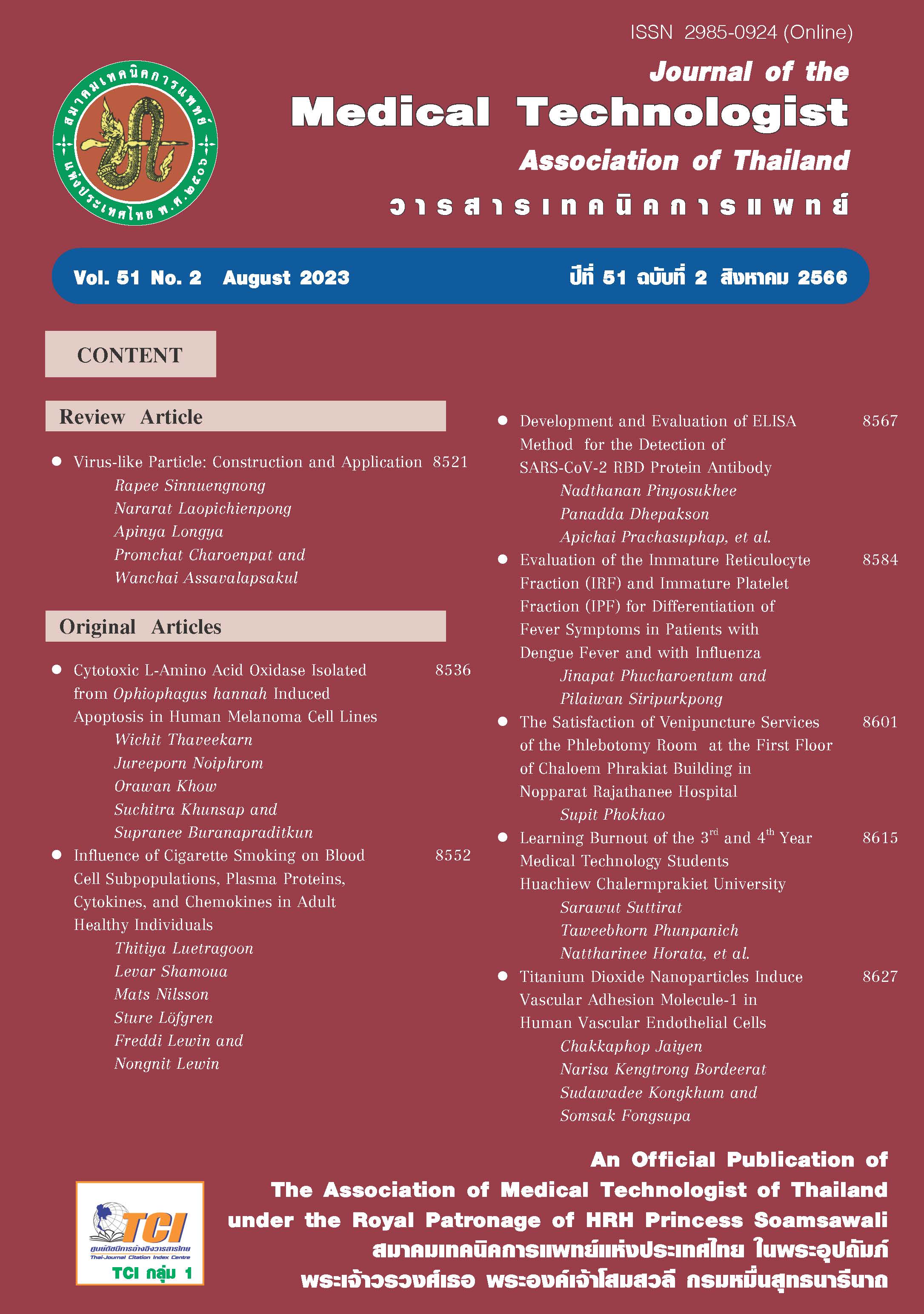
อนุภาคนาโนไทเทเนียมไดออกไซด์กระตุ้นการแสดงออกของโปรตีนยึดเกาะในเซลล์บุผนังหลอดเลือด
บทคัดย่อ
วัสดุนาโนเป็นวัสดุที่มีอนุภาคนาโนเป็นส่วนประกอบ มีความเสถียรสูง และทนต่อการกัดกร่อน ปัจจุบันมีการนำอนุภาคนาโนมาใช้ประโยชน์ในการผลิตสินค้าอุตสาหกรรมและสินค้าอุปโภคและบริโภคมากขึ้น ทำให้มีโอกาสสัมผัสและนำเข้าสู่ร่างกายมากขึ้น โดยผ่านทางการหายใจ การกิน และทางผิวหนัง หลังจากนั้นอนุภาคนาโนจะเข้าสู่ระบบไหลเวียนโลหิตผ่านเซลล์บุผนังหลอดเลือดและอาจทำให้เกิดพิษต่อเซลล์ได้ อนุภาคนาโนไทเทเนียมไดออกไซด์เป็นอนุภาคนาโนที่มีการนำมาใช้ผลิตสินค้าหลายชนิด ได้แก่ ผลิตภัณฑ์สีทาบ้าน ผลิตภัณฑ์ป้องกันการติดเชื้อ เครื่องสำอาง ครีมกันแดด ยารักษาโรค เครื่องมือแพทย์ ผลิตภัณฑ์อาหาร และสารกำจัดแมลง ดังนั้นการวิจัยนี้จึงมีวัตถุประสงค์เพื่อศึกษาความเป็นพิษ และผลกระทบโดยตรงของอนุภาคนาโนไทเทเนียมไดออกไซด์ต่อเซลล์บุผนังหลอดเลือด โดยใช้เซลล์เพาะเลี้ยงชนิด HMEC-1 วัดปริมาณการเข้าสู่เซลล์ของอนุภาคนาโนไทเทเนียมไดออกไซด์ อัตราการมีชีวิตของเซลล์ การสร้างอนุมูลอิสระ และการแสดงออกของโมเลกุลยึดเกาะบนผิวเซลล์ ผลการศึกษาพบว่าที่ความเข้มข้น 50-1,000 มิลลิกรัมต่อลิตร อนุภาคนาโนไทเทเนียมไดออกไซด์เข้าสู่เซลล์ได้เฉลี่ยร้อยละ 15.4 ของปริมาณที่สัมผัส มีผลเพิ่มอัตราการมีชีวิตของเซลล์ เพิ่มระดับสารอนุมูลอิสระในเซลล์ และกระตุ้นการแสดงออกของโปรตีน VCAM-1 อย่างมีนัยสำคัญ (p<0.05) สรุปว่า อนุภาคนาโนไทเทเนียมไดออกไซด์มีผลกระตุ้นการแสดงออกของโปรตีนยึดเกาะ VCAM-1 ซึ่งอาจนำไปสู่กระบวนการอักเสบของเซลล์บุผนังหลอดเลือด และภาวะหลอดเลือดแดงแข็งได้
เอกสารอ้างอิง
Vongpoon N, Pradipachitti N, Puvanakijjakorn P, Sukniwatchai K, Keskovit S, Poomkam J. Current regulation on nanoproducts of Thai Food and Drug Administration. Thai Pharmaceutical and Health Science Journal 2015;10(3):132-8. (in Thai)
Buxton DB, Lee SC, Wickline SA, Ferrari M. National Heart, Lung and Blood Nanotechnology Working Group. Recommendations of The National Heart, Lung and Blood Institute Nanotechnology Working Group, Circulation. 2003; 108 (22): 2737-42.
Peters A, Wichmann HE, Tuch T, Heinrich J, Heyder J. Respiratory effects are associated with the number of ultrafine particles. Am. J. Respir. Crit. Care Med 1997; 155: 1376-83.
Donaldson K, Tran L, Jimenez L, et al. Combustion-derived nanoparticles: a review of their toxicology following inhalation exposure. Particle Fibre Toxicol 2005; 2: 1-14.
Hogg JC, Timens W. The pathology of chronic obstructive pulmonary disease. Annu Rev Pathol 2009; 4: 435-59.
Lu D, Luo Q, Chen R, et al. Chemical multi-fingerprinting of exogenous ultrafine particle in human serum and pleural effusion. Nat. Commun 2020; 11: 2567-74.
Calderón-Garcidueas L, González-Maciel A, Mukherjee PS, et al. Combustion-and friction-derived magnetic air pollution nanoparticle in human heart. Environ. Res [internet]. 2019 Sep [cited 2020 Mach 10];176. Available from: https://www.sciencedirect.com/science/article/abs/pii/S0013935119303640 via%3Dihub
Miiller MR, Raftis JB, Langrish JP, et al. Inhaled nanoparticles accumulate at sites of vascular disease. ACS Nano 2017; 11: 4542-52.
Kido T, Tamagawa E, Bai N. Particulate matter induces translocation of LI-6 from the lung to the systemic circulation. Am J Respir Cell Mol Biol 2021; 44 (2): 197-204.
Miyata R, Bai N, Vincent R, Sin DD, Eeden SF. Statin reduce ambient particle matter-induced lung inflammation by promoting the clearance of particulate matter < 10 µm from lung tissues. Chest 2013; 143 (2): 452-60.
Foster CA. VCAM-1/alpha 4-integrin adhesion pathway: therapeutic target for allergic inflammatory disorders. J Allergy Clin Immunol. 1996;98: S270-7.
Ross R. Atherosclerosis--an inflammatory disease. N Engl J Med. 1999 Jan 14;340 (2):115-26.
Ballantyne CM, Nambi V. Markers of inflammation and their clinical significance. Atheroscler Suppl. 2005;6(2):21-9.
Lusis AJ. Atherosclerosis. Nature 2000; 407: 233-41.
Joppa P, Petrasova D, Stancak B, Tkacova R. Systemic inflammation in patient with COPD and pulmonary hypertension. Chest 2006; 130: 326-33.
Laratta CR, Eeden SV. Acute exacerbation of chronic obstructive pulmonary disease: cardiovascular links. BioMed Research International 2014; 528789: 1-18.
Danesh J, Whincup P, Walker M, et al. Low-grade inflammation and coronary heart disease: prospective study and updated meta-analysis. Br Med J 2000; 321: 199-204.
Cargnello M, Gordon TR, Murray CB. Solution-phase synthesis of titanium dioxide nanoparticles and nanocrystals. Chem. Rev. 2014; 114: 9319-45.
Braun JH, Baidins A, Marganski RE. TiO2 pigment technology: A review. Prog. Org. Coat.1992; 20: 105–38.
Shand M, Anderson AJ. Aqueous phase photocatalytic nitrate destruction using titania based materials: Routes to enhanced performance and prospects for visible light activation. Catal. Sci. Technol. 2013; 3: 879–99.
Gopinath KP, Madhav NV, Krishnan A, Malolan R, Rangarajan G. Present applications of titanium dioxide for the photocatalytic removal of pollutants from water: A review. J. Environ. Manag. [internet]. 2020 Jun [cited 2021 Oct 21]; 270. Available from: https://www.Sciencedirect.com /science/article/pii/S0301479720308367via%3Dihub
Racovita AD. Titanium Dioxide: Structure, Impact, and Toxicity. Int. J. Environ. Res. Public Health [internet]. 2022 May [cited 2022 Oct 22];19. Available from: https://www.mdpi.com/journal/ijerph
Kreyling WG, Holzwarth U, Schleh C, et al. Quantitative biokinetics over a 28day period of freshly generated, pristine, 20 nm titanium dioxide nanoparticle aerosols in healthy adult rats after a single two-hour inhalation exposure. Particle and Fibre Toxicology 2019.16:1-29.
Qia Y, Weia S, Xin T, et al. Passage of exogenous fine particles from the lung into the brain in humans and animals.PNAS 2022; 119 (26):1-9.
Wolfgang G, Kreyling WG, Holzwarth U, et al. Quantitative biokinetics over a 28 days period of freshly generated, pristine, 20 nm titanium dioxide nanoparticle aerosols in healthy adult rats after a single two-hour inhalation exposure. Particle Fibre Toxicol 2019; 16: 29–56.
Schleicher N, Norra S, Chai F, Chen Y, Wang S, Steuben D. Anthropogenic versus geogenic contribution to total suspended atmospheric particulate matter and its variations during a two-year sampling period in Beijing China. J. Environ. Monit. 2010; 12: 434–41
Shakeel M, Jabeen F, Shabbir S, Asghar MS, Khan MS, Chaudhry AS. Toxicity of nano-titanium dioxide (TiO2-NP) through various routes of exposure: A review. Biol. Trace Elem. Res 2016; 172: 1–36.
Shi H, Magaye R, Castranova V, Zhao J. Titanium dioxide nanoparticles: a review of current toxicological
data. Part Fibre Toxicol 2013; 10:15–16.
Yah CS, Simate GS, Iyuke SE. Nanoparticles toxicity and their routes of exposures. Pak J Pharm Sci 2012;
: 477- 91.
Arora S, Rajwadw JM, Paknikar K. Nanotoxicology and in vitro studies: the need of hour. Appl Pharm 2010; 258: 151-65.
Bermudez E, Mangum JB, Wong BA, et al. Pulmonary responses of
mice, rats, and hamsters to sub-chronic inhalation of ultrafine titanium dioxide particles. Toxicol Sci 2004;
(2): 347–57.
Lindberg HK, Falck GCM, Catalán J, et al. Genotoxicity of inhaled
nanosized TiO2 in mice. Mutat Res Genn Toxicol Environ Mutagen 2012; 745(1–2): 58-64.
LeBlanc AJ, Moseley AM, Chen BT, Frazer D, Castranova V, Nurkiewicz TR. Nanoparticle inhalation impairs
coronary microvascular reactivity via a local reactive oxygen species dependent mechanism. Cardiovasc
Toxicol 2010; 10(1): 27–36.
Nurkiewicz TR, Porter DW, Hubbs AF, et al. Pulmonary particulate
matter and systemic microvascular dysfunction. Res Rep Health Eff Inst 2011; 164: 3-48.
Prichard E. Inorganic analytes: Determination, In: Trace analysis: A structured approach to obtaining reliable results. 1st Ed. Cambridge: Crown copyright for the Laboratory of the Government Chemist by the Royal Society of Chemistry 1996; 95-152.
Sayes CM, Reed KL, Warheit DB. Assessing toxicity of fine and nanoparticles: comparing in vitro measurements to in vivo pulmonary toxicity profiles. Toxicol Sci 2007; 97: 163-80.
Warheit DB, Hoke RA, Finlay C, Donner EM, Reed KL, Sates CM. Development of a base set of toxicity tests using ultrafine TiO2 particles as a component of nanoparticle risk management. Toxicol Lett 2007; 171: 99-110.
Folkmann JK, Risom L, Jacobsen NR, Wallin H, Loft S, Moller P. Oxidatively damaged DNA in rats exposed by oral gavage to C60 fullerenes and single-walled carbon nanotubes. Environ Health Perspect 2009; 117: 703-8.
Karlsson HL, Cronholm P, Gustafsson J, Moller L. Copper oxide nanoparticles are highly toxic: a comparison between metal oxide nanoparticles and carbon nanotubes. Chem Res Toxicol 2008; 21: 1726-32.
Foster CA. VCAM-1/alpha 4-integrin adhesion pathway: therapeutic target for allergic inflammatory disorders. J Allergy Clin Immunol 1996; 98: 270-7.
Nia Y, Millour S, Noël L, Krystek P, Jong WD, Guérin T. Determination of Ti from TiO2 Nanoparticles in Biological Materials by Different ICP-MS Instruments: Method Validation and Applications. J Nanomed Nanotechnol 2015; 6 (2): 1-8.
Aranda A, Sequedo L, Tolosa L, et al. Dichloro-dihydro-fluorescein
diacetate (DCFH-DA) assay: A quantitative method for oxidative stress assessment of nanoparticle-treated cells 2013; Toxicol Vitro 27 (2013) 954-63
Bradford MM. A rapid and sensitive method for the quantitation of microgram quantities of protein utilizing the principle of protein-dye binding. Anal Biochem 1976; 72: 248-54.
Sukwong P, Kongseng S, Chaicherd S, Yoovathaworn K, Tubtimkuna S, Pissuwan D. Comparison effects of titanium dioxide nanoparticles on immune cells in adaptive and innate immune system. IET Nanobiotechnol 2017; 11: 759-65.
Peng L, Barczak AJ, Barbeau RA, et al. Whole Genome Expression Analysis Reveals Differential Effects of TiO2 Nanotubes on Vascular Cells. Nano Lett 2010;10(1):143-8.
Montiel-Dávalos A, Ventura-Gallegos JL, Alfaro-Moreno E, et al. TiO₂ nanoparticles induce dysfunction and activation of human endothelial cells. Chem Res Toxicol 2012; 25(4): 920-30.
Kido T, Tamagawa E, Bai N, et al. Particulate matter induces translocation of IL-6 from the lung to the systemic circulation. Am J Respir Cell Mol Biol. 2011; 44(2): 197-204.
Miyata R, Bai N, Vincent R, Sin DD, Van Eeden SF. Statins reduce ambient particulate matter-induced lung inflammation by promoting the clearance of particulate matter, < 10 µm from lung tissues. Chest. 2013; 143(2): 452-60.
Miyata R, Hiraiwa K, Cheng JC, et al. Statins attenuate the development of atherosclerosis and endothelial dysfunction induced by exposure to urban particulate matter (PM10). Toxicol Appl Pharmacol 2013; 272(1): 1-11.
Shukla RK, Sharma V, Pandey AK, Singh S, Sultana S, Dhawan A, ROS-mediated genotoxicity induced by titanium dioxide nanoparticles in human epidermal cells. Toxicol Vitro 2011; 25: 231–41
Nel A, Xia T, Ma¨dler L, Li N. Toxic Potential of Materials at the Nano level. Science 2006; 311: 622-7.
Han SG, Newsome B, Hennig B. Titanium dioxide nanoparticles increase inflammatory responses in vascular endothelial cells. Toxicology 2013; 122: 1-8.
Gurr JR, Wang AS, Chen CH, Jan KY. Ultrafine titanium dioxide particles in the absence of photoactivation can induce oxidative damage to human bronchial epithelial cells. Toxicology 2005; 213(1–2): 66-73.
Long TC, Tajuba J, Sama P, et al. Nanosize titanium dioxide stimulates reactive oxygen species in brain microglia and damages neurons in vitro. Environ Health Perspect 2007; 115(11): 1631-7.
Skocaj M, Filipic M, Petkovic J, Novak S. Titanium dioxide in our everyday life; Is it safe?. Radiol Oncol 2011; 45(4): 227–47.
Eeden SF, Sin DD. Chronic obstructive pulmonary disease: A chronic systemic inflammatory disease. Respiration 2008; 75: 224–38.
Deem TL, Cook-Mills JM. Vascular cell adhesion molecule 1 (VCAM-1) activation of endothelial cell matrix metalloproteinases: role of reactive oxygen species, Blood. 2004;104(8): 2385–93.
Eeden SF, Sin DD. Chronic obstructive pulmonary disease: A chronic systemic inflammatory disease. Respiration 2008; 75: 224–38.
Sin DD, Man SF. Chronic obstruction pulmonary disease: a novel risk factor for cardiovascular disease. Can J Physiol Pharmacol 2005; 83: 8-13.
ดาวน์โหลด
เผยแพร่แล้ว
รูปแบบการอ้างอิง
ฉบับ
ประเภทบทความ
สัญญาอนุญาต
ลิขสิทธิ์ (c) 2023 วารสารเทคนิคการแพทย์

อนุญาตภายใต้เงื่อนไข Creative Commons Attribution-NonCommercial-NoDerivatives 4.0 International License.


