
Direction-Dependent Soft Tissue Thickness in Facial Asymmetry: A Three-Dimensional Analysis Using Facial Scanning and Cone Beam Computed Tomography
Article Sidebar
Main Article Content
Abstract
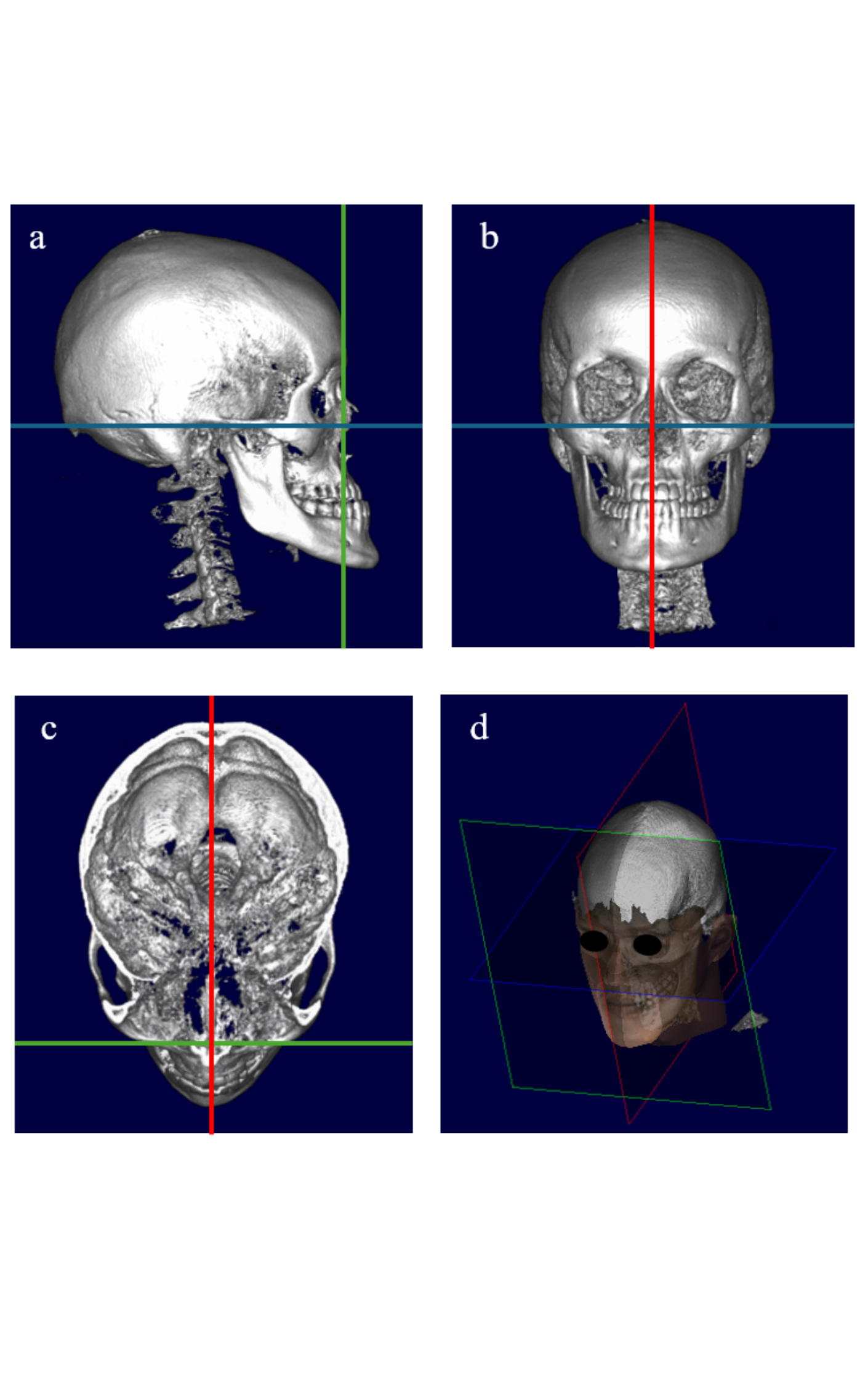
Background: Facial asymmetry is a common craniofacial condition affecting facial aesthetics and function. Although skeletal discrepancies are the primary determinant, the role of direction-dependent soft tissue thickness in shaping facial morphology remains unclear. Objective: To investigate directiondependent patterns of soft tissue thickness in patients with facial asymmetry using integrated three-dimensional facial scanning and cone beam computed tomography. Materials and methods: This cross-sectional study included 60 adults aged 18–35 years undergoing orthodontic and orthognathic evaluation. Cone beam computed tomography and three-dimensional facial scan datasets were fused to construct composite craniofacial models. Bilateral skeletal landmarks were identified, and soft tissue thickness was measured relative to the midsagittal plane in transverse and sagittal directions. Paired statistical tests compared measurements between deviated and contralateral sides. Results: Significant skeletal asymmetry was observed at multiple craniofacial landmarks, particularly at the antegonial notch (2.09 ± 4.40 mm), orbitale (1.66 ± 3.16 mm), and anterior ramus notch (1.34 ± 1.94 mm), among others (P < 0.01). Direction-dependent patterns of soft tissue thickness was identified. In the transverse, soft tissue was generally thinner on the deviated side at the anterior ramus notch, canini, antegonial notch, and gonion (P < 0.01). In contrast, sagittal measurements showed greater soft tissue thickness on the deviated side at the condylion, antegonial notch, and gonion (P < 0.01). Conclusion: Soft tissue thickness in facial asymmetry demonstrates direction-dependent patterns and only partially reflects skeletal deviations. Integrated three-dimensional imaging may improve evaluation of skeletal–soft tissue relationships and assist orthodontic and orthognathic treatment planning.
Article Details

This work is licensed under a Creative Commons Attribution-NonCommercial-NoDerivatives 4.0 International License.
References
Thiesen G, Gribel BF, Freitas MP. Facial asymmetry: a current review. Dent Press J Orthod 2015;20:110-25.
Cheong YW, Lo LJ. Facial asymmetry: etiology, evaluation, and management. Chang Gung Med J 2011;34:341-51.
Zhuang J, Ma H, Wang C, Kong X, Chen Y, Su X, et al. Applying 3D scanning to evaluate facial symmetry in Asian populations. J Plast Reconstr Aesthet Surg 2024;99:11-7.
Jearanai T, Samruajbenjakun B, Chanmanee P. Relationship between bilateral landmarks of facial asymmetry in skeletal Class II and Class III in vertical dimension: 3D facial scan and cone-beam computed tomography. Diagnostics (Basel) 2024;14:590.
Zhu Y, Zhao Y, Wang Y. A review of three-dimensional facial asymmetry analysis methods. Symmetry 2022;14:1414.
Kim HJ, Noh HK, Park HS. Differences in facial soft tissue deviations in Class III patients with different types of mandibular asymmetry: a cone-beam computed tomography study. Korean J Orthod 2023;53:402-19.
Li J, Wu S, Mei L, Wen J, Marra J, Lei L, et al. Facial asymmetry of the hard and soft tissues in skeletal Class I, II, and III patients. Sci Rep 2024;14:4966.
Hwang HS, Yuan D, Jeong KH, Uhm GS, Cho JH, Yoon SJ. Three-dimensional soft tissue analysis for the evaluation of facial asymmetry in normal occlusion individuals. Korean J Orthod 2012;42:56-63.
Lee MS, Chung DH, Lee JW, Cha KS. Assessing soft-tissue characteristics of facial asymmetry with photographs. Am J Orthod Dentofacial Orthop 2010;138:23-31.
Rustemeyer J, Martin A. Soft tissue response in orthognathic surgery patients treated by bimaxillary osteotomy: cephalometry compared with two-dimensional photogrammetry. Oral Maxillofac Surg 2013;17:33-41.
Choi KY. Analysis of facial asymmetry. Arch Craniofac Surg 2015;16:1-10.
Nur RB, Çakan DG, Arun T. Evaluation of facial hard and soft tissue asymmetry using cone-beam computed tomography. Am J Orthod Dentofacial Orthop 2016;149:225-37.
Lee JY, Han SH, Ryu HS, Lee HM, Kim SC. Cone-beam computed tomography analysis of transverse dental compensation in patients with skeletal Class III malocclusion and facial asymmetry. Korean J Orthod 2018;48:357-66.
Elkenawy I, Fijany L, Colak O, Paredes NA, Gargoum A, Abedini S, et al. An assessment of the magnitude, parallelism, and asymmetry of micro-implant-assisted rapid maxillary expansion in non-growing patients. Prog Orthod 2020;21:42.
Cantarella D, Dominguez-Mompell R, Mallya SM, Moschik C, Pan HC, Miller J, et al. Changes in the midpalatal and pterygopalatine sutures induced by micro-implantsupported skeletal expander analyzed with a novel 3D method based on CBCT imaging. Prog Orthod 2017;18:34.
Muñoz SRT, Cantin M, Perez-Rojas F, Suazo I. Evaluation of facial asymmetry using soft-tissue thickness for forensic purposes. Int J Morphol 2011;29:1033-9.
Tam TKM, Guo R, Liu H, Lin Y. Hard and soft tissue asymmetry in patients with skeletal Class III malocclusion: a cone-beam computed tomography study. Diagnostics (Basel) 2023;13:869.
Setvaji NR, Muthuswamy Pandian S. Evaluation of soft tissue compensations in subjects with facial asymmetry using cone-beam computed tomography: a retrospective study. J Pharm Bioallied Sci 2024;16(Suppl 1):S120-4.
Ajmera DH, Singh P, Leung YY, Gu M. Three-dimensional evaluation of soft-tissue response to osseous movement after orthognathic surgery in patients with facial asymmetry: a systematic review. J Oral Maxillofac Surg 2021;79:1230-40.
Wu Z, Gao X, Long H, Lai W. Quantitative analysis of facial symmetry using three-dimensional technology. J Craniofac Surg 2022;33:e345-50.
Vasconcelos BC, Gonçalves FA, Andrade A, Guillen M, Landim F. Mandibular asymmetry: literature review and case report. J Oral Maxillofac Surg 2012;70:e95-102.
Siqueira de Lima L, Brunetto DP, da Cunha Gonçalves Nojima M. Evaluation of facial soft tissue thickness in symmetric and asymmetric subjects using cone-beam computed tomography. Angle Orthod 2019;89:799-806.
Fan Y, Zhang Y, Chen G, He W, Song G, Matthews H, et al. Automated assessment of mandibular shape asymmetry in three dimensions. Sci Rep 2022;12:14521.
Meundi MA, David CM. Application of cone beam computed tomography in facial soft tissue thickness measurements for craniofacial reconstruction. J Forensic Dent Sci 2019;11:7-12.
Ho C-T, Lin H-H, Liou EJW, Lo L-J. Three-dimensional surgical simulation improves the planning for correction of facial prognathism and asymmetry: a qualitative and quantitative study. Sci Rep 2017;7:40423.
Lyu L, Zhang MJ, Wen AN, Wang S, Zhao YJ, Yong W, et al. 3D facial mask for facial asymmetry diagnosis. Comput Methods Programs Biomed 2024;248:108089.
Hsu P-J, Denadai R, Pai BCJ, Lin H-H, Lo L-J. Outcome of facial contour asymmetry after conventional two-dimensional versus computer-assisted three-dimensional planning in cleft orthognathic surgery. Sci Rep 2020;10:2346.
Wermker K, Kleinheinz J, Jung S, Dirksen D. Soft tissue response and facial symmetry after orthognathic surgery. J Craniomaxillofac Surg 2014;42:1540-6.
Jokić D, Uglešić V, Macan D, Knežević P. Soft tissue changes after mandibular setback and bimaxillary surgery in Class III patients. J Craniomaxillofac Surg 2013;41:e65-70.
Da Pozzo F, Gibelli D, Beltramini GA, Dolci C, Giannì AB, Sforza C. The effect of orthognathic surgery on soft-tissue facial asymmetry: a longitudinal three-dimensional analysis. J Craniomaxillofac Surg 2020;48:1120-6.
