
Comparison of Convolutional Neural Networks Architectures for Screening Mandibular-Plane-to-Hyoid Distance as a Risk Indicator of Obstructive Sleep Apnea on Lateral Cephalograms
Article Sidebar
Main Article Content
Abstract
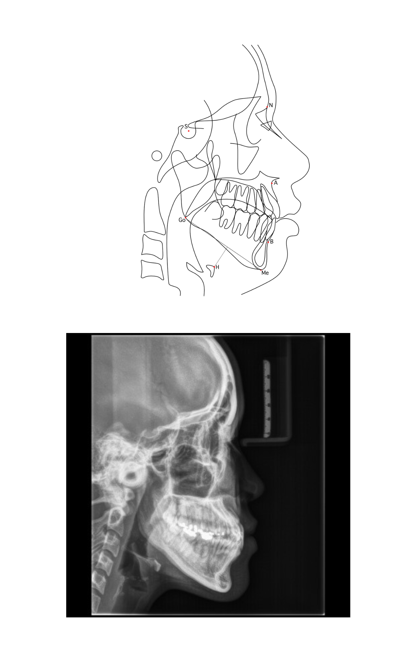
Background: Lateral cephalometric radiographs can aid in screening obstructive sleep apnea (OSA) through the mandibular-plane-to-hyoid (MP-H) distance. An automated tool based on this measure could support OSA screening. However, the accuracy of such tools depends on the performance of AI-assisted models, making it essential to evaluate and compare their effectiveness in detecting MP-H distance for reliable clinical application. Objective: To evaluate the performance of different convolutional neural network (CNN) architectures in classifying patients into short and long MP-H groups. Materials and methods: A total of 304 pre-orthodontic lateral cephalometric radiographs from adults (age 18-56 years) were classified into short MP-H (< 18 mm) and long MP-H (≥ 18 mm) groups. Four CNN architectures (DenseNet-121, ResNet-50, EfficientNet-B0, and MobileNetV3) were trained to classify short and long MP-H groups. To address class imbalance, weighted binary cross entropy loss functions (weights ranging from 1 to 5) were applied, assigning greater penalties to misclassification of the minority class. Results: In the scenario without application of weighted cross entropy loss, DenseNet-121 achieved the overall high screening performance, with sensitivity = 0.87, specificity = 0.95, precision = 0.79, F1-score = 0.82, accuracy = 0.93, AUROC = 0.91, and AUPRC = 0.83. MobileNetV3 consistently demonstrated the lowest performance. Weighted loss functions provided inconsistent benefits across architectures. DenseNet-121 showed consistent performance among all weights. Conclusion: DenseNet-121 shows potential of screening long MP-H distance from lateral cephalograms. Weighted loss functions may provide improvements, but model selection remains the more critical factor.
Article Details

This work is licensed under a Creative Commons Attribution-NonCommercial-NoDerivatives 4.0 International License.
References
Spicuzza L, Caruso D, Di Maria G. Obstructive sleep apnoea syndrome and its management. Ther Adv Chronic Dis 2015;6(5):273-85.
Neruntarat C, Chantapant S. Prevalence of sleep apnea in HRH Princess Maha Chakri Srinthorn Medical Center, Thailand. Sleep Breath 2011;15(4):641-8.
Alansari RA. The role of orthodontics in management of obstructive sleep apnea. Saudi Dent J 2022;34(3):194-201.
Ryu HH, Kim CH, Cheon SM, Bae WY, Kim SH, Koo SK, et al. The usefulness of cephalometric measurement as a diagnostic tool for obstructive sleep apnea syndrome: a retrospective study. Oral Surg Oral Med Oral Pathol Oral Radiol 2015;119(1):20-31.
deBerry-Borowiecki B, Kukwa A, Blanks RH. Cephalometric analysis for diagnosis and treatment of obstructive sleep apnea. Laryngoscope 1988;98(2):226-34.
Neelapu BC, Kharbanda OP, Sardana HK, Balachandran R, Sardana V, Kapoor P, et al. Craniofacial and upper airway morphology in adult obstructive sleep apnea patients: a systematic review and meta-analysis of cephalometric studies. Sleep Med Rev 2017;31:79-90.
Auvenshine Rc Dds P, Pettit Nj Dmd MSD. The hyoid bone: an overview. Cranio 2020;38(1):6-14.
Bailey EF. Activities of human genioglossus motor units. Respir Physiol Neurobiol 2011;179(1):14-22.
Banhiran W, Wanichakorntrakul P, Metheetrairut C, Chiewvit P, Planuphap W. Lateral cephalometric analysis and the risks of moderate to severe obstructive sleepdisordered breathing in Thai patients. Sleep Breath 2013;17(4):1249-55.
Jo JH, Park JW, Jang JH, Chung JW. Hyoid bone position as an indicator of severe obstructive sleep apnea. BMC Pulm Med 2022;22(1):349.
Kazmouz S, Calzadilla N, Choudhary A, McGinn LS, Seaman A, Purnell CA. Radiographic findings predictive of obstructive sleep apnea in adults: a systematic review and meta-analysis. J Craniomaxillofac Surg 2025;53(2):162-80.
Riley R, Guilleminault C, Herran J, Powell N. Cephalometric analyses and flow-volume loops in obstructive sleep apnea patients. Sleep 1983;6(4):303-11.
Ahmadi K, Amali A, Saedi B, Dasdar S, Rashedi S, Kianfar N, et al. Evaluation of cephalometric indices in patients with obstructive sleep apnea in comparison with healthy individuals. Adv Oral Maxillofac Surg 2022;5:100250.
Andersson L, Brattström V. Cephalometric analysis of permanently snoring patients with and without obstructive sleep apnea syndrome. Int J Oral Maxillofac Surg 1991; 20(3):159-62.
Yoon HJ, Kim DR, Gwon E, Kim N, Baek SH, Ahn HW, et al. Fully automated identification of cephalometric landmarks for upper airway assessment using cascaded convolutional neural networks. Eur J Orthod 2022;44(1): 66-77.
Manakitsa N, Maraslidis GS, Moysis L, Fragulis GF. A review of machine learning and deep learning for object detection, semantic segmentation, and human action recognition in machine and robotic vision. Technologies 2024;12(2):15.
Russell BC, Torralba A, Murphy KP, Freeman WT. LabelMe: a database and web-based tool for image annotation. Int J Comput Vis 2008;77(1):157-73.
Jeong Y, Nang Y, Zhao Z. Automated evaluation of upper airway obstruction based on deep learning. Biomed Res Int 2023;2023:8231425.
Jeong HG, Kim T, Hong JE, Kim HJ, Yun SY, Kim S, et al. Automated deep neural network analysis of lateral cephalogram data can aid in detecting obstructive sleep apnea. J Clin Sleep Med 2023;19(2):327-37.
Huang G, Liu Z, Van Der Maaten L, Weinberger KQ. Densely connected convolutional networks. In: Proceedings of the IEEE Conference on Computer Vision and Pattern Recognition (CVPR); 2017 Jul 21-26; Honolulu, HI, USA. p. 4700-8.
He K, Zhang X, Ren S, Sun J. Deep residual learning for image recognition. In: Proceedings of the IEEE Conference on Computer Vision and Pattern Recognition (CVPR); 2016 Jun 27-30; Las Vegas, NV, USA. p. 770-8.
Tan M, Le QV. EfficientNet: rethinking model scaling for convolutional neural networks [Internet]. arXiv; 2019 [cited 2025 Aug 7]. Available from: https://arxiv.org/ abs/1905.11946.
Howard A, Sandler M, Chu G, Chen L-C, Chen B, Tan M, et al. Searching for MobileNetV3. In: Proceedings of the IEEE/ CVF International Conference on Computer Vision (ICCV); 2019 Oct 27 - Nov 2; Seoul, South Korea. p. 1314-24.
Tsuiki S, Nagaoka T, Fukuda T, Sakamoto Y, Almeida FR, Nakayama H, et al. Machine learning for image-based detection of patients with obstructive sleep apnea: an exploratory study. Sleep Breath 2021;25(4):2297-305.
Özdemir Ö, Sönmez EB. Weighted cross-entropy for unbalanced data with application on COVID X-ray images. In: 2020 Innovations in Intelligent Systems and Applications Conference (ASYU); 2020 Oct 15-17; Istanbul, Turkey. p. 1-5.
de Hond AAH, Steyerberg EW, van Calster B. Interpreting area under the receiver operating characteristic curve. Lancet Digit Health 2022;4(12):e853-5.
Ozenne B, Subtil F, Maucort-Boulch D. The precision–recall curve overcame the optimism of the receiver operating characteristic curve in rare diseases. J Clin Epidemiol 2015;68(8):855-9.
Koo TK, Li MY. A guideline of selecting and reporting intraclass correlation coefficients for reliability research. J Chiropr Med 2016;15(2):155-63.
Goceri E. Medical image data augmentation: techniques, comparisons and interpretations. Artif Intell Rev 2023:1-45.
Goyal M, Mahmoud QH. A systematic review of synthetic data generation techniques using generative AI. Electronics 2024;13(17):3509.
