
Differences between Posteroanterior Cephalometric Analysis By 2D Conventional Posteroanterior Cephalograms and 3D Models Generated from Cone Beam Computed Tomography
Article Sidebar
Main Article Content
Abstract
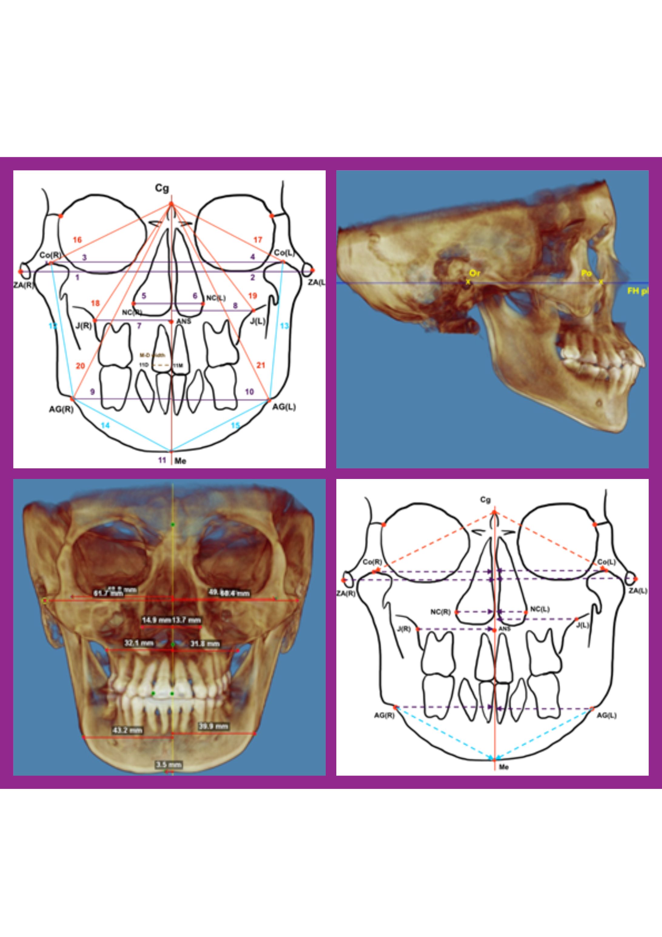
Background: This study compared the differences in posteroanterior (PA) cephalometric analysis on a two-dimensional (2D)-PA cephalogram with cone beam computed tomography (CBCT) via Dolphin imaging software®. Materials and methods: Retrospective data from 35 patients who required orthodontic treatment (35 2D-PA cephalograms and 35 CBCT images) were obtained. All radiographs were imported into the Dolphin imaging program®, aligned, and calibrated for magnification using patients’ tooth sizes derived from dental models. Landmarks were identified, and linear measurements modified from Grummons analysis were evaluated. 2D-PA cephalograms and CBCT measurements were compared via paired t tests (P < 0.05). Results: According to Grummon PA cephalometric analysis, significant differences (P < 0.05) were observed in 10 horizontal, 2 vertical, and 2 mandibular length variables between 2D-PA cephalograms and CBCT. Conclusion: Compared with CBCT, 2D-PA cephalography could acceptably indicate the degree of menton deviation. However, the measurements above the maxillary area from 2D-PA cephalograms are significantly different from those from CBCT. PA cephalograms could be used as an initial tool to evaluate lower facial asymmetry. However, for cases requiring detailed analysis and comprehensive planning, CBCT might be necessary.
Article Details

This work is licensed under a Creative Commons Attribution-NonCommercial-NoDerivatives 4.0 International License.
References
Athanasiou AE. Orthodontic cephalometry. London: Mosby-Wolfe; 1995.p.9-11
Proffit WR, Fields HW, Sarver DM. Contemporary orthodontics. 5th ed: Elsevier Health Sciences; 2014:147-275.
Dindaroglu F, Yetkiner E. Cone beam computed tomography in orthodontics. Turk J Orthod 2016;29(1):16-21.
Abdelkarim A. Cone-beam computed tomography in orthodontics. Dent J (Basel) 2019;7(3):89
Premkumar S. Textbook of orthodontics. New Delhi: Elsevier; 2015:253-313.
Venkatesh E, Elluru SV. Cone beam computed tomography: basics and applications in dentistry. J Istanb Univ Fac Dent 2017;51(3 Suppl 1):S102-21.
Meena R, Chauhan R, Bharvada K. A review on cone beam computed tomography in orthodontics. SVOA Dent 2022;3(1):47-51.
Bajaj K, Rathee P, Jain P, Panwar VR. Comparison of the reliability of anatomic landmarks based on PA cephalometric radiographs and 3D CT scans in patients with facial asymmetry. Int J Clin Pediatr Dent 2011;4(3):213-23.
Hechler SL. Cone-beam CT: applications in orthodontics. Dent Clin North Am. 2008;52(4):809-23.
Kaygisiz E, Tortop T. Cone beam computed tomography in orthodontics. In: Halefoglu AM, Editor.Computed Tomography - Advanced Applications. intechopen 2017.p.117-38.
Leonardi R, Annunziata A, Caltabiano M. Landmark identification error in posteroanterior cephalometric radiography: a systematic review. Angle Orthod 2008;78(4):761-5.
Sicurezza E, Greco M, Giordano D, Maiorana F, Leonardi R. Accuracy of landmark identification on postero-anterior cephalograms. Prog Orthod 2012;13(2):132-40.
Damstra J, Fourie Z, Ren Y. Evaluation and comparison of postero-anterior cephalograms and cone-beam computed tomography images for the detection of mandibular asymmetry. Eur J Orthod 2013;35(1):45-50.
Yousefi F, Rafiei E, Mahdian M, Mollabashi V, Saboonchi SS, Hosseini SM. Comparison efficiency of posteroanterior cephalometry and cone-beam computed tomography in detecting craniofacial asymmetry: a systematic review. Contemp Clin Dent 2019;10(2):358-71.
Sfogliano L, Abood A, Viana G, Kusnoto B. Cephalometric evaluation of posteroanterior projection of reconstructed three-dimensional Cone beam computed tomography, two-dimensional conventional radiography, and direct measurements. J World Fed Orthod 2016;5(1):22-7.
Chen C-M, Tseng Y-C, Ko EC, Chen M, Chen K-J, Cheng J-H. Comparisons of Jaw Line and Face Line after Mandibular Setback: Intraoral Vertical Ramus versus Sagittal Split Ramus Osteotomies. BioMed Res Int 2018;2018:1-7.
Kang H-J, Kim J-R, Kim Y-I. Validity of Horizontal Reference Planes on Cone-Beam Computed Tomography Generated Postero-Anterior Cephalogram. Maxillofac Plast Reconstr Surg 2011;33(4):346-51.
Hwang HS, Hwang CH, Lee KH, Kang BC. Maxillofacial 3-dimensional image analysis for the diagnosis of facial asymmetry. Am J Orthod Dentofacial Orthop 2006;130(6):779-85.
Szuhanek C, Nagib R, Sabo-Meze A, Buzatu R, Malita D. Cephalometric antero-posterior parameter evaluation in orthodontic patients with facial asymmetries. J Dent Oral Implants 2020;2(3):8-15.
Edgren B. The frontal cephalometric analysis – the forgotten perspective. Orthod Pract US 2013;4(5):28-33.
Garib DG, Calil LR, Leal CR, Janson G. Is there a consensus for CBCT use in orthodontics? Dental Press J Orthod 2014;19(5):136-49.
Tai B, Goonewardene MS, Murray K, Koong B, Islam SMS. The reliability of using postero-anterior cephalometry and cone-beam CT to determine transverse dimensions in clinical practice. Aust Orthod J 2014;30(2):132-42.
Patterson Dental Holdings, Inc. Dolphin Imaging: Comprehensive Software Guide. 2020 Available from: https://www.dolphinimaging.com/product/ThreeD.
Wang CH, Randazzo L. Evolution of imaging and management systems in orthodontics. Am J Orthod Dentofacial Orthop 2016;149(6):798-805.
Scarfe WC, Pinheiro LR, Farman AG. CBCT and the orthodontic patient: Cone-beam computed tomography offers key benefits in all phases of orthodontics-from diagnosis to post-treatment. Dimensions of Dental Hygiene 2014;12(8):18-21.
