
The Relationship between Skeletal Configuration and Soft Tissue Changes af ter Bracket Debonding using Repeatable Photographic Tool
Article Sidebar
Main Article Content
Abstract
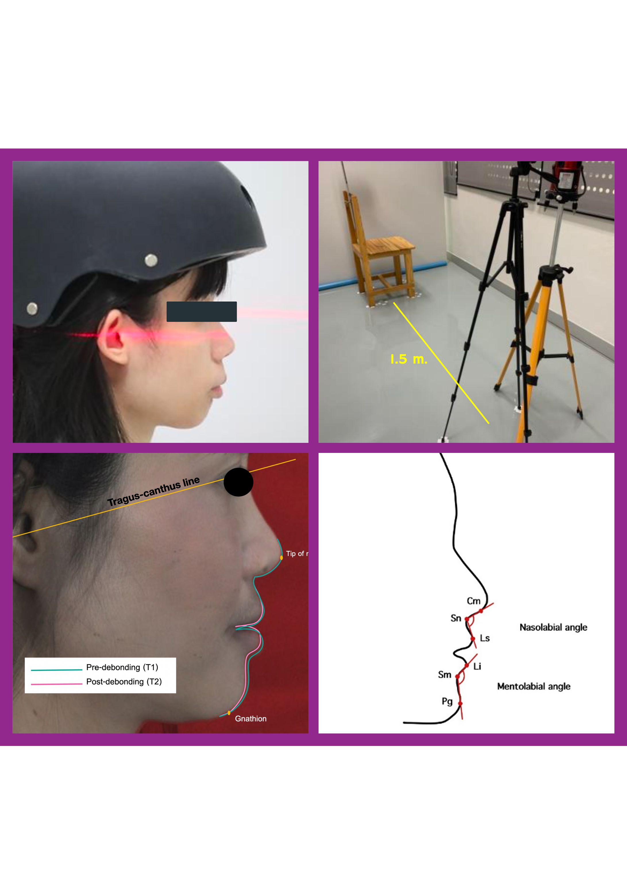
Background: The presence of labial orthodontic appliances may impact final esthetic change after debonding. The skeletal configurations that support the soft tissue profile have not been examined their impact on the lip profile after debonding. Objective: To evaluate the effect of bonded orthodontic brackets on the lip change after the debonding and determine the correlation between the change in the lip profile and skeletal configuration. Materials and methods: Photographs were taken with a head fixer in thirty-three patients who had completed fixed orthodontic treatment before and immediately after bracket debonding to investigate the results of the change in the nasolabial and mentolabial angles using the Paired t test ( = 0.05). The posttreatment lateral cephalometric measurements were used to find the correlation of skeletal configuration to the change in soft tissue profile using Pearson’s correlation and one-way ANOVA. Results: Mentolabial angle significantly increased after debonding (P = 0.04). However, the Pearson correlation between soft tissue changes and underlying skeletal configurations was insignificant. (SNA with nasolabial angle: r = 0.13, P = 0.46; SNB with mentolabial angle: r = - 0.00, P = 0.98). Using one-way ANOVA, skeletal configurations demonstrated no significant difference compared with the mean difference in nasolabial angle (P = 0.69) and mean difference in mentolabial angle (P = 0.15). Conclusion: After debonding, the lower lip profile was flattened, however, the upper lip profile was maintained compared with the nose. There was no significant correlation between the change of nasolabial/mentolabial angles and the skeletal configurations.
Article Details

This work is licensed under a Creative Commons Attribution-NonCommercial-NoDerivatives 4.0 International License.
References
Ackerman JL, Proffit WR, Sarver DM. The emerging soft tissue paradigm in orthodontic diagnosis and treatment planning. Clin Orthod Res 1999;2(2):49-52.
Abed Y, Har-Zion G, Redlich M. Lip posture following debonding of labial appliances based on conventional profile photographs. Angle Orthod 2009;79(2):235-9.
Fernández-Riveiro P, Smyth-Chamosa E, Suárez-Quintanilla D, Suárez-Cunqueiro M. Angular photogrammetric analysis of the soft tissue facial profile. Eur J Orthod 2003;25(4):393-9.
Milosević SA, Varga ML, Slaj M. Analysis of the soft tissue facial profile of Croatians using of linear measurements. J Craniofac Surg 2008;19(1):251-8.
Ahn HW, Chang YJ, Kim KA, Joo SH, Park YG, Park KH. Measurement of three-dimensional perioral soft tissue changes in dentoalveolar protrusion patients after orthodontic treatment using a structured light scanner. Angle Orthod 2014;84(5):795-802.
Eidson L, Cevidanes LH, de Paula LK, Hershey HG, Welch G, Rossouw PE. Three-dimensional evaluation of changes in lip position from before to after orthodontic appliance removal. Am J Orthod Dentofacial Orthop 2012;142(3):410-8.
Jang KS, Bayome M, Park JH, Park KH, Moon HB, Kook YA. A three-dimensional photogrammetric analysis of the facial esthetics of the Miss Korea pageant contestants. Korean J Orthod 2017;47(2):87-99.
Jeon H, Lee SJ, Kim TW, Donatelli RE. Three-dimensional analysis of lip and perioral soft tissue changes after debonding of labial brackets. Orthod Craniofac Res 2013;16(2):65-74.
Kim YK, Lee NK, Moon SW, Jang MJ, Kim HS, Yun PY. Evaluation of soft tissue changes around the lips after bracket debonding using three-dimensional stereophotogrammetry. Angle Orthod 2015;85(5):833-40.
Hayashida H, Ioi H, Nakata S, Takahashi I, Counts AL. Effects of retraction of anterior teeth and initial soft tissue variables on lip changes in Japanese adults. Eur J Orthod 2011;33(4):419-26.
Farias Gomes A, Moreira DD, Zanon MF, Groppo FC, Haiter-Neto F, Freitas DQ. Soft tissue thickness in Brazilian adults of different skeletal classes and facial types: a cone beam CT - Study. Leg Med (Tokyo) 2020;47:101743.
Proffit WR, Fields HW, Sarver DM. Contemporary orthodontics 6th ed. Mosby Elsevier 2018.
Arnett GW, Bergman RT. A soft tissue cephalometric analysis. Am J Orthod Dentofacial Orthop 1993;104(4):348-66.
Ackerman JL, Proffit WR. Soft tissue considerations in orthodontic treatment. Angle Orthod 1997;67(1):9-20.
Sarver DM. The importance of soft tissue in the assessment of the orthodontic patient. World J Orthod 2015;16(1):4-19.
Evans JD. Straightforward statistics for the behavioral sciences. Brooks/Cole Publishing 1996.
Moshkelgosha V, Fathinejad S, Pakizeh Z, Shamsa M, Golkari A. Photographic facial soft tissue analysis by means of linear and angular measurements in an adolescent Persian population. Open Dent J 2015;9:346-56.
Esmaeili S, Mohammadi NM, Khosravani S, Eslamian L, Motamedian SR. Evaluation of facial profile characteristics of aesthetically pleasing Iranian faces. J World Fed Orthod 2023;12(2):76-89.
Penna V, Fricke A, Iblher N, Eisenhardt SU, Stark GB. The attractive lip: a photomorphometric analysis. J Plast Reconstr Aesthet Surg 2015;68(7):920-9.
Sarnoff DS, Gotkin RH. Six steps to the “perfect” lip. J Drugs Dermatol 2012;11(9):1081-8.
Seeholzer C, Klug A. Patients’ perception of soft tissue changes after orthodontic treatment. J Orofac Orthop 2019;80:47-56.
