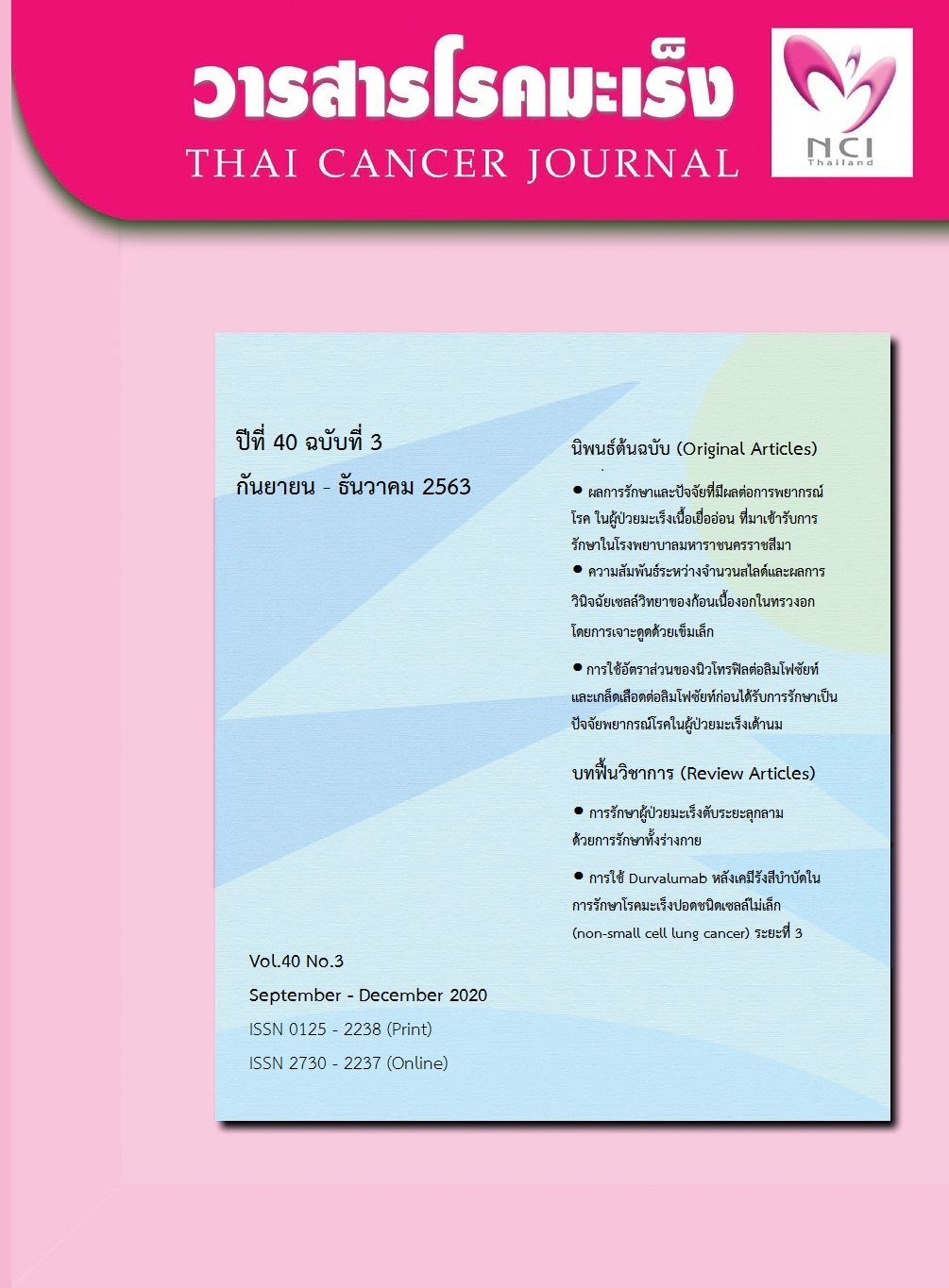
ความสัมพันธ์ระหว่างจำนวนสไลด์และผลการวินิจฉัยเซลล์วิทยาของก้อนเนื้องอก ในทรวงอกโดยการเจาะดูดด้วยเข็มเล็ก
คำสำคัญ:
การเจาะดูดด้วยเข็มเล็ก สไลด์ FNA การตรวจวินิจฉัยชิ้นเนื้อ ก้อนเนื้อที่ปอดบทคัดย่อ
การเจาะดูดด้วยเข็มเล็ก (FNA) เป็นวิธีการหนึ่งที่แพทย์นิยมใช้สำหรับตรวจวินิจฉัยเบื้องต้นในติ่งหรือก้อนเนื้อในส่วนต่าง ๆ ของร่างกายรวมทั้งอวัยวะในช่องทรวงอก เนื่องจากทำได้ง่าย ผู้ป่วยมีอาการแทรกซ้อนภายหลังการทำหัตถการน้อย การเก็บชิ้นเนื้อทำได้โดยการใช้เข็มปราศจากเชื้อขนาดเล็กแทงไปยังรอยโรคที่ปอดซึ่งอาจนำร่องด้วยการเอ็กซเรย์คอมพิวเตอร์หรือคลื่นเสียงสะท้อน แล้วเจาะดูดเซลล์มาป้ายบนแผ่นสไลด์เพื่อส่งตรวจวินิจฉัยทางเซลล์วิทยา อย่างไรก็ตามพบว่าสไลด์ FNA มีจำนวนไม่แน่นอน ตั้งแต่ 1 ถึง 16 สไลด์ต่อราย ซึ่งปริมาณสไลด์ที่ต่างกันอาจส่งผลต่อการวินิจฉัยของพยาธิแพทย์ การศึกษานี้จึง มีวัตถุประสงค์เพื่อศึกษาความสัมพันธ์ระหว่างจำนวนสไลด์ FNA และผลการวินิจฉัยเซลล์วิทยาของก้อนเนื้องอกในทรวงอก โดยศึกษาแบบย้อนหลังจากข้อมูลผู้ป่วยของสถาบันโรคทรวงอก จำนวน 3782 ราย ที่มารับการตรวจระหว่างปี พ.ศ. 2554 ถึง 2562 ผลการศึกษาพบว่า การส่งสไลด์จำนวนมากขึ้น มีแนวโน้มในการได้ผลวินิจฉัยเพิ่มขึ้น ทั้งนี้จำนวนสไลด์ตั้งแต่ 4 แผ่นขึ้นไป มีอัตราการได้ผลการวินิจฉัยสูงกว่าการไม่ได้ผลการวินิจฉัย (P <0.001) ขณะที่จำนวนการส่งมากกว่า 6 สไลด์ไม่พบความแตกต่างของผลการวินิจฉัย โดยสรุปผลการวินิจฉัย FNA มีความสัมพันธ์กับปริมาณสไลด์ ซึ่งปริมาณที่ควรเตรียม อาจอยู่ระหว่าง 4-6 สไลด์ต่อครั้ง ขณะเดียวกันการส่งมากกว่า 6 สไลด์ขึ้นไป อาจถือว่าเกินความจำเป็น
เอกสารอ้างอิง
media market company; 2012. p. 10-4.
2. Layfield LJ, Esebua M, Dodd L, Giorgadze T, Schmidt RL. The Papanicolaou Society of Cytopathology guidelines for respiratory cytology:
Re-producibility of categories among observers. Available from : https://pubmed.ncbi.nlm.nih. gov30294354. Accessed September 15, 2020
3.สมจินต์ จินดาวิจักษณ์, วิษณุ ปานจันทร์, อาคม วีระวัฒนะ, วีรวุฒิ อิ่มสำราญ. การตรวจทางเซลล์วิทยาด้วยเข็มขนาดเล็กกรณีที่เป็นโรคก้อนของต่อมไทรอยด์
ชนิดหลายก้อน (Multinodular goiter). แนวทางการตรวจวินิจฉัยและรักษาโรคมะเร็งต่อมไทรอยด์. กรุงเทพฯ: บริษัทโฆสิตการพิมพ์ จำกัด; 2558. หน้า 17-8.
4.นิลยา สุคำวัง. หัตถการเจาะดูดด้วยเข็มเล็ก (Fine needle aspiration) ของก้อนที่เต้านม สำหรับแพทย์และบุคลากรทางการแพทย์. วารสารเทคนิคการแพทย์เชียงใหม่ 2554;44:89-92.
5.Luz LP, Moreira DM, Khan M, Eloubeidi MA. Predictors of malignancy in EUS-guided FNA for mediastinal lymphadenopathy
in patients without history of lung cancer. Ann Thorac Med 2011;6:126-30.
6. Gangopadhyay M, Chakrabarti I, Ghosh N, Giri A. Computed tomography guided fine needle aspiration cytology of mass
lesions of lung: Our experience. Indian J Med Paediatr Oncol;32:192-6.
7. Barta JA,Henschke CI,Flores RM,Yip R, Yanke levitz DF, Powell CA. Lung cancer diagnosis by fine needle aspiration is associated
with reduction in resection of nonmalignant lung nodules. Ann Thorac Surg2017;103:1795-801.
8. Itonaga M, Yasukawa S, Shimokawa T, Takenaka M, Fukutake N, Ogura T, et al. Comparison of 22 G standard and Franseen
needles in endoscopic ultrasound-guided fine-needle aspiration for diagnosing pancreatic mass lesions: Study protocol for a
controlled trial. Trials;20:816.
9. Fassina A, Corradin M, Zardo D, Cappellesso R, Corbetti F, Fassan M. Role and accuracy of rapid
on-site evaluation of CT-guided fine needle aspiration cytology of lung nodules. Cytopathol 2011;22:306-12.
10. Shrestha MK, Ghartimagar D, Ghosh A. Computed tomogram guided fine-needle aspiration cytology of lung and mediastinal masses with
cytological correlation: a study of 257 cases in Western region of Nepal. Nepal Med Coll J 2014;16:80-3.
11. Aj L, Kalra N, Bhatia A, Srinivasan R, Gulati A, Kapoor R, et al. Fusion image-guided and ultrasound-guided fine needle aspiration in patients with
suspected hepatic metastases. J Clin Exp Hepatol 2019;9:547-53.
12.Eltoum IA, Chhieng DC, Jhala D, Jhala NC, Crowe DR, Varadarajulu S, et al. Cumulative sum procedure in evaluation of EUS-guided FNA cytology:
the learning curve and diagnostic performance beyond sensitivity and specificity. Cytopathology 2007;18:143-50.
13. Manucha V, Kaur G, Verma K. Endoscopic ultrasound-guided fine needle aspiration (EUS-FNA) of mediastinal lymph nodes:
experience from region with high prevalence of tuberculosis. Diagn Cytopathol 2013;41:1019-22.
14.สมบูรณ์ ศีลาวัฒน์. ปัญหาและทางแก้ของการให้การวินิจฉัยโรคในงานบริการของ fine-needle aspiration cytology. Chula Med J 2544;45:276-82.
15. Shidham VB, Varsegi GM, D'Amore K, Shidham A. Preparation and using phantom lesions to practice fine needle aspiration biopsies.
Available from: https://www.jove.com/t/1404.Accessed September 21,2020 .
16.พรสุดา จิตรกสิกร, นิลทิตา ศรีไพบูลย์กิจ. สถิติผู้ป่วยโรคมะเร็งปอด. หน่วยทะเบียนมะเร็ง คณะแพทยศาสตร์โรงพยาบาลรามาธิบดี; 2557
เข้าถึงจาก https://med.mahidol.ac.th/cancercenter/th/news/event/22082016-1833th.สืบค้นเมื่อวันที่ 17 ตุลาคม 2564
17. Department of Pathology. สิ่งส่งตรวจจากการทำ Fine needle aspiration (FNA). Chiang Mai:Department of Pathology,
Faculty of Medicine, Chiangmai University; 2020 Available from:https://w1med.cmu.ac.th/patho/FNA.html.Accessed August 3, 2020.
18.Chaiwun B, Sukhamwang N, Lekawanvijit S, Sukapan K, Rangdaeng S, Muttarak M, et al. Atypical and suspicious categories in fine
needle aspiration cytology of the breast: histological and mammographical correlation and clinical significance. Singapore Med J 2005;46:706-9.
19. Sinha SK, Chatterjee M, Bhattacharya S, Pathak SK, Mitra RB, Karak K, et al. Diagnostic evaluation of extrapulmonary tubercu-losis by fineneedle
aspiraton (FNA) supple-mented with AFB smear and culture. J Indian Med Assoc 2003101:588,90-1.
20. Zhang Y, Gomez-Fernandez CR, Jorda M, Ganjei-Azar P. Fine-needle aspiration (FNA) and pleural fluid cytology diagnosis of benign metastasizing
pleomorphic adenoma of the parotid gland in the lung: a case report and review of literature. Diagn Cytopathol 2009; 37:828-31.
21.Kothari K, Tummidi S, Agnihotri M, Sathe P, Naik L. This 'Rose' Has no Thorns-Diagnostic Utility of 'Rapid On-Site Evaluation' (ROSE)
in Fine Needle Aspiration Cytology. Indian J Surg Oncol 2019; 10:688-98.
22.Lee JC, Kim H, Kim HW, Lee J, Paik KH, Kang J, et al. It is necessary to exam bottom and top slide smears of EUS-FNA for pancreastic cancer.
Hepatobiliary Pancreat Dis Int 2018 ;17:553-8.
ดาวน์โหลด
เผยแพร่แล้ว
ฉบับ
ประเภทบทความ
สัญญาอนุญาต
บทความทีตีพิมพ์ในวารสารโรคมะเร็งนี้ถือว่าเป็นลิขสิทธิ์ของมูลนิธิสถาบันมะเร็งแห่งชาติ และผลงานวิชาการหรือวิจัยของคณะผู้เขียน ไม่ใช่ความคิดเห็นของบรรณาธิการหรือผู้จัดทํา


