
Stone culturing: a more effective approach to diagnosing bacterial infections in kidney stone formers
Article Sidebar
Main Article Content
Abstract
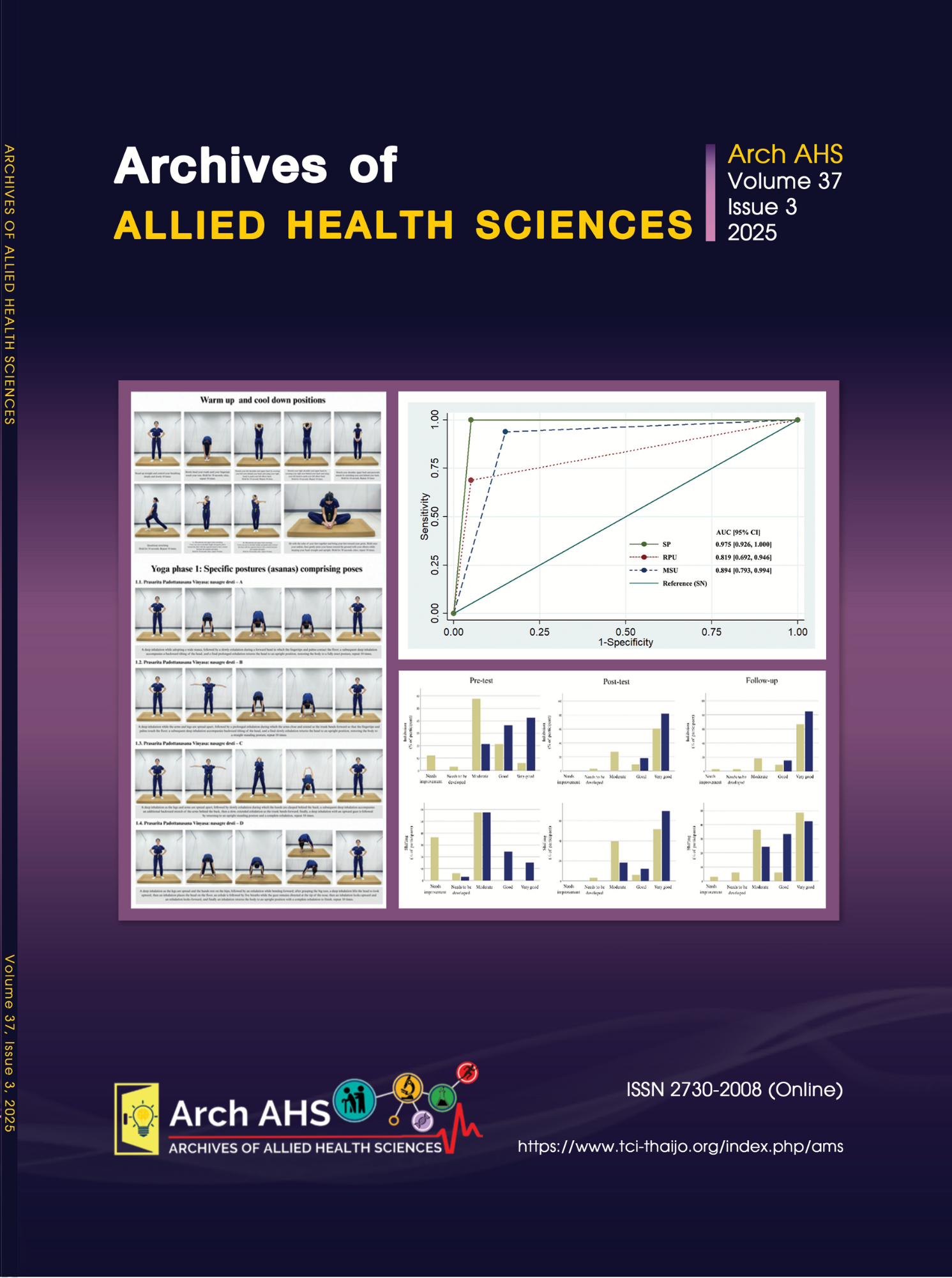
According to the bacteria found in stone niduses, these bacteria may be responsible for lithogenesis. Therefore, we considered culturing stone niduses (SN) as the gold standard for comparing bacterial culture results from stone peripheries (SP), renal pelvic urine (RPU), and midstream urine (MSU), including an evaluation of performance. Data from 36 kidney stone formers were collected, including demographics, imaging diagnostics, urinalysis, and preoperative midstream urine culture. The samples of SN, SP, and RPU were cultured to identify microorganisms. SN were also analyzed for their chemical composition. Diagnostic testing, including sensitivity, specificity, positive likelihood ratio (LR+), negative likelihood ratio (LR-), positive predictive value (PPV), negative predictive value (NPV), and area under the curve (AUC) with 95% confidence intervals (CI), was performed. The results showed that 16 (44.44%) SN, 17 (47.22%) SP, 12 (33.33%) RPU, and 18 (50.00%) MSU were positive for bacterial culture. For the performance testing that compares SN and the other three specimens, the sensitivity, LR+, PPV, NPV, and AUC of SP culture (sensitivity = 100%, LR+ = 20.00, PPV = 94.10%, NPV = 100%, AUC = 0.975) demonstrated a high level, exceeding that of RPU and MSU cultures. The level of agreement between SN and SP cultures was almost perfect (0.94). Escherichia coli, Klebsiella pneumoniae, and Proteus mirabilis were the most commonly isolated bacteria from stone and urine cultures. Moreover, P. mirabilis and E. coli were the most common bacteria isolated from struvite and calcium oxalate monohydrate (COM) stone compositions, respectively. Our data indicate that culturing SN exhibited higher concordance with SP than the urine culture. P. mirabilis and E. coli were the most commonly isolated from infection-induced (i.e., struvite) and non infection-induced (i.e., COM) stones, respectively. Integrating stone and urine cultures into the diagnostic workflow for bacterial infections in KSFs is recommended.
Article Details

This work is licensed under a Creative Commons Attribution-NonCommercial-NoDerivatives 4.0 International License.
References
Schwaderer AL, Wolfe AJ. The association between bacteria and urinary stones. Ann Transl Med 2017; 5(2): 32.
Ripa F, Pietropaolo A, Montanari E, Hameed BMZ, Gauhar V, Somani BK. Association of kidney stones and recurrent UTIs: the chicken and egg situation. A systematic review of literature. Curr Urol Rep 2022; 23(9): 165-74.
Razi A, Ghiaei A, Dolatabadi FK, Haghighi R. Unraveling the association of bacteria and urinary stones in patients with urolithiasis: an update review article. Front Med (Lausanne) 2024; 11: 1401808.
Miano R, Germani S, Vespasiani G. Stones and urinary tract infections. Urol Int 2007; 79 (suppl 1): 32-6.
European Association of Urology. Urolithiasis [online] 2025 [cited 2025 May 9]. Available from: https://uroweb.org/guidelines/ urolithiasis.
Oliver R, Ghosh A, Geraghty R, Moore S, Somani BK. Successful ureteroscopy for kidney stone disease leads to resolution of urinary tract infections: Prospective outcomes with a 12-month follow-up. Cent European J Urol 2017; 70(4): 418-23.
Tavichakorntrakool R, Prasongwattana V, Sungkeeree S, Saisud P, Sribenjalux P, Pimratana C, et al. Extensive characterizations of bacteria isolated from catheterized urine and stone matrices in patients with nephrolithiasis. Nephrol Dial Transplant 2012; 27(11): 4125-30.
Tavichakorntrakool R, Boonsiri P, Prasongwatana V, Lulitanond A, Wongkham C, Thongboonkerd V. Differential colony size, cell length, and cellular proteome of Escherichia coli isolated from urine vs. stone nidus of kidney stone patients. Clin Chim Acta 2017; 466: 112-9.
Mariappan P, Loong CW. Midstream urine culture and sensitivity test is a poor predictor of infected urine proximal to the obstructing ureteral stone or infected stones: a prospective clinical study. J Urol 2004; 171(6 Pt 1): 2142-5.
Paonessa JE, Gnessin E, Bhojani N, Williams JC Jr, Lingeman JE. Preoperative bladder urine culture as a predictor of intraoperative stone culture results: clinical implications and relationship to stone composition. J Urol 2016; 196(3): 769-74.
Mariappan P, Smith G, Bariol SV, Moussa SA, Tolley DA. Stone and pelvic urine culture and sensitivity are better than bladder urine as predictors of urosepsis following percutaneous nephrolithotomy: a prospective clinical study. J Urol 2005; 173(5): 1610-4.
Liu M, Chen J, Gao M, Zeng H, Cui Y, Zhu Z, Chen H. Preoperative midstream urine cultures vs renal pelvic urine culture or stone culture in predicting systemic inflammatory response syndrome and urosepsis after percutaneous nephrolithotomy: a systematic review and meta-analysis. J Endourol 2021; 35(10): 1467-78.
Procop GW, Church DL, Hall GS, Janda WM, Koneman EW, Schreckenberger PC, editors Color atlas and textbook of diagnostic microbiology. 7th ed. Wolters Kluwer Health; 2017.
Uttamamul N, Jitpean S, Lulitanond A, Wonglakorn L, Sae-Ung N, Boonsiri P, et al. Risk factors for canine magnesium ammonium phosphate urolithiasis associated with bacterial infection. J Vet Sci 2021; 23(1): e6.
Daudon M, Petay M, Vimont S, Deniset A, Tielens F, Haymann JP, et al. Urinary tract infection inducing stones: some clinical and chemical data. Comptes Rendus Chimie 2022; 25: 315-34.
McHugh ML. Interrater reliability: the kappa statistic. Biochem Med (Zagreb) 2012; 22(3): 276-82.
Chewcharat A, Curhan G. Trends in the prevalence of kidney stones in the United States from 2007 to 2016. Urolithiasis 2021; 49(1): 27-39.
Stamatelou K, Goldfarb DS. Epidemiology of kidney stones. Healthcare (Basel) 2023; 11(3): 424.
Sritippayawan S, Borvornpadungkitti S, Paemanee A, Predanon C, Susaengrat W, Chuawattana D, et al. Evidence suggesting a genetic contribution to kidney stone in northeastern Thai population. Urol Res 2009; 37(3): 141-6.
Ferraro PM, Taylor EN, Curhan GC. Factors associated with sex differences in the risk of kidney stones. Nephrol Dial Transplant 2023; 38(1): 177-83.
Shang W, Li Y, Ren Y, Yang Y, Li H, Dong J. Nephrolithiasis and risk of hypertension: a meta-analysis of observational studies. BMC Nephrol 2017; 18(1): 344.
Taylor EN, Stampfer MJ, Curhan GC. Diabetes mellitus and the risk of nephrolithiasis. Kidney Int 2005; 68(3): 1230-5.
Daudon M, Doré JC, Jungers P, Lacour B. Changes in stone composition according to age and gender of patients: a multivariate epidemiological approach. Urol Res 2004; 32(3): 241-7.
De Lorenzis E, Boeri L, Gallioli A, Fontana M, Zanetti SP, Longo F, et al. Feasibility and relevance of urine culture during stone fragmentation in patients undergoing percutaneous nephrolithotomy and retrograde intrarenal surgery: a prospective study. World J Urol 2021; 39(6): 1725-32.
Shah P, Baral R, Agrawal CS, Lamsal M, Baral D, Khanal B. Urinary Calculi: A microbiological and biochemical analysis at a tertiary care hospital in eastern Nepal. Int J Microbiol 2020; 2020: 8880403.
Das P, Gupta G, Velu V, Awasthi R, Dua K, Malipeddi H. Formation of struvite urinary stones and approaches towards the inhibition-A review. Biomed Pharmacother 2017; 96: 361-70.
Diri A, Diri B. Management of staghorn renal stones. Ren Fail 2018; 40(1): 357-62.
Chutipongtanate S, Sutthimethakorn S, Chiangjong W, Thongboonkerd V. Bacteria can promote calcium oxalate crystal growth and aggregation. J Biol Inorg Chem 2013; 18(3): 299-308.
Barr-Beare E, Saxena V, Hilt EE, Thomas-White K, Schober M, Li B, et al. The interaction between Enterobacteriaceae and calcium oxalate deposits. PLoS One 2015; 10(10): e0139575.
Amimanan P, Tavichakorntrakool R, Fong-Ngern K, Sribenjalux P, Lulitanond A, Prasongwatana V, et al. Elongation factor Tu on Escherichia coli isolated from urine of kidney stone patients promotes calcium oxalate crystal growth and aggregation. Sci Rep 2017; 7(1): 2953.
Kanlaya R, Naruepantawart O, Thongboonkerd V. Flagellum is responsible for promoting effects of viable Escherichia coli on calcium oxalate crystallization, crystal growth, and crystal aggregation. Front Microbiol 2019; 10: 2507.
