
Automatic delineation of organs at risk for prostate cancer on computed tomography images using convolutional neural networks
Article Sidebar
Main Article Content
Abstract
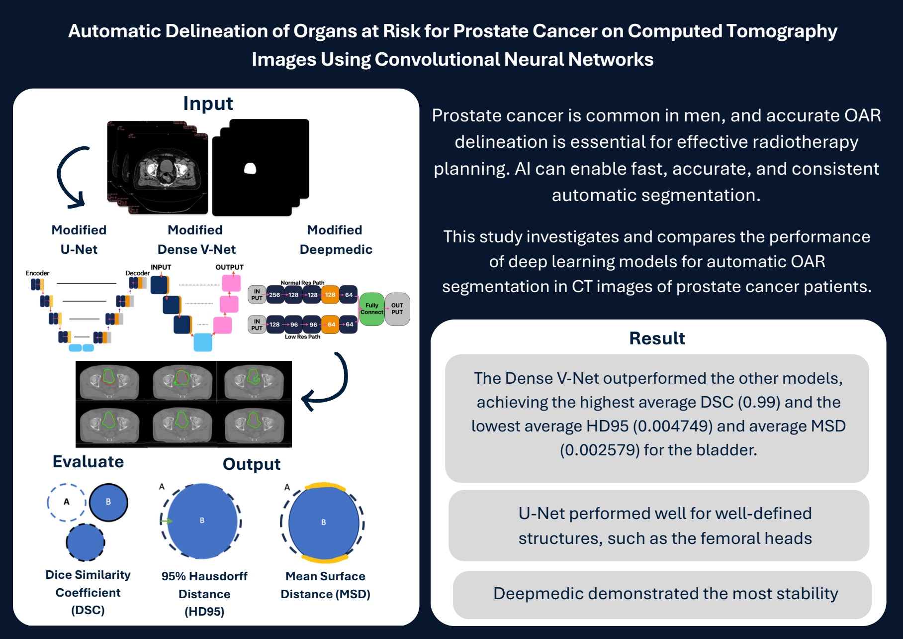
Background: Prostate cancer is the fourth most common cancer worldwide and the second most common in men. Accurate identification of organs at risk (OARs) is crucial for improving treatment planning, making the process faster and more efficient. Artificial intelligence has become a vital tool in medicine, reducing delays and improving treatment workflow by enabling precise, consistent automatic organ delineation.
Objectives: This study investigates and compares the performance of deep learning models for automatic OAR segmentation in CT images of prostate cancer patients.
Materials and methods: Using 2,011 CT images from the Prostate-Anatomical-Edge-Cases dataset, the U-Net, Dense V-Net, and DeepMedic models were evaluated under the same parameters for segmenting the bladder, rectum, and left and right femoral heads. Performance was assessed using the Dice Similarity Coefficient (DSC), 95th percentile Hausdorff Distance (HD95), and Mean Surface Distance (MSD).
Results: The Dense V-Net outperformed the other models, achieving the highest DSC (0.99) and the lowest HD95 (0.00) and MSD (0.00) for the bladder. U-Net performed well for well-defined structures, such as the femoral heads, while DeepMedic demonstrated the most stability.
Conclusion: Dense V-Net’s superior performance highlights its effectiveness in medical image segmentation, particularly in prostate cancer treatment.
Article Details

This work is licensed under a Creative Commons Attribution-NonCommercial-NoDerivatives 4.0 International License.
Personal views expressed by the contributors in their articles are not necessarily those of the Journal of Associated Medical Sciences, Faculty of Associated Medical Sciences, Chiang Mai University.
References
Bott SR, Ng KL. Prostate cancer. Brisbane: Exon Publications; 2021.
World Cancer Research Fund International. Global cancer incidence in men [Internet]. 2020 [cited 2023 May 2]. Available from: www.wcrf.org/cancertrends/worldwide-cancer-data/.
Zou KH, Warfield SK, Bharatha A, Tempany CM, Kaus MR, Kikinis R, et al. Statistical validation of image segmentation quality based on a spatial overlap index. Acad Radiol. 2004; 11(2): 178-89. doi: 10.1016/S1076-6332(03)00671-8.
Vinod SK, Jameson MG, Min M, Holloway LC. Uncertainties in volume delineation in radiation oncology: a systematic review and recommendations for future studies. Radiother Oncol. 2016; 121(2): 169-79. doi: 10.1016/j.radonc.2016.09.009.
Wong J, Huang V, Wells D, Giambattista J, Otto K, Alexander A, et al. Implementation of deep learningbased auto-segmentation for radiotherapy planning structures: a workflow study at two cancer centers. Radiat Oncol. 2021; 16(1): 101. doi: 10.1186/ s13014-021-01831-4.
Wang J, Chen Y, Xie H, Luo L, Tang Q. Evaluation of auto-segmentation for EBRT planning structures using deep learning-based workflow on cervical cancer. Sci Rep. 2022; 12(1): 13650. doi: 10.1038/s41598-022-18084-0.
Tataru OS, Vartolomei MD, Rassweiler JJ, Virgil O, Lucarelli G, Ferro M, et al. Artificial intelligence and machine learning in prostate cancer patient management-current trends and future perspectives. Diagnostics (Basel). 2021; 11(2): 354. doi: 10.3390/diagnostics11020354.
Sultana S, Robinson AR, Song DY, Lee J. Automatic multi-organ segmentation in computed tomography images using hierarchical convolutional neural network. J Med Imaging (Bellingham). 2020; 7(5): 055001. doi: 10.1117/1.JMI.7.5.055001.
Rouvière O, Moldovan PC, Vlachomitrou A, Gouttard S, Riche B, Rabilloud M, et al. Combined modelbased and deep learning-based automated 3D zonal segmentation of the prostate on T2-weighted MR images: clinical evaluation. Eur Radiol. 2022; 32(5): 3248-59. doi: 10.1007/s00330-021-08408-5.
Polymeri E, Johnsson ÅA, Enqvist O, Ulén J, Pettersson N, Kjölhede H, et al. Artificial intelligencebased organ delineation for radiation treatment planning of prostate cancer on computed tomography. Adv Radiat Oncol. 2024; 9(3): 101383. doi: 10.1016/j.adro.2023.101383.
Russell SJ, Norvig P. Artificial intelligence: a modern approach. 4th ed. Hoboken: Pearson; 2021.
Mehralivand S, Yang D, Harmon SA, Xu D, Xu Z, Turkbey B, et al. Deep learning-based artificial intelligence for prostate cancer detection at biparametric MRI. Abdom Radiol (NY). 2022; 47(4): 1425-34. doi: 10.1007/s00261-022-03419-2.
Isaksson LJ, Pepa M, Zaffaroni M, Marvaso G, Alterio D, Jereczek-Fossa BA, et al. Machine learningbased models for prediction of toxicity outcomes in radiotherapy. Front Oncol. 2020; 10: 790. doi: 10.3389/fonc.2020.00790.
Isaksson LJ, Pepa M, Summers P, Zaffaroni M, Vincini MG, Jereczek-Fossa BA, et al. Comparison of automated segmentation techniques for magnetic resonance images of the prostate. BMC Med Imaging. 2023; 23(1): 32. doi: 10.1186/s12880-023-00974-y.
Hirashima H, Nakamura M, Baillehache P, Fujimoto Y, Nakagawa S, Mizowaki T, et al. Development of in-house fully residual deep convolutional neural network-based segmentation software for the male pelvic CT. Radiat Oncol. 2021; 16(1): 135. doi: 10.1186/ s13014-021-01867-6.
Elguindi S, Zelefsky MJ, Jiang J, Veeraraghavan H, Deasy JO, Hunt MA, et al. Deep learning-based auto-segmentation of targets and organs-at-risk for magnetic resonance imaging only planning of prostate radiotherapy. Phys Imaging Radiat Oncol. 2019; 12: 80-6. doi: 10.1016/j.phro.2019.11.006.
Ronneberger O, Fischer P, Brox T. U-Net: convolutional networks for biomedical image segmentation. In: Navab N, Hornegger J, Wells WM, Frangi AF, editors. Medical Image Computing and Computer-Assisted Intervention - MICCAI 2015. Cham: Springer; 2015. p. 234-41.
Lempart M, Nilsson MP, Scherman J, Gustafsson CJ, Nilsson M, Olsson LE, et al. Pelvic U-Net: multilabel semantic segmentation of pelvic organs at risk for radiation therapy anal cancer patients using a deeply supervised shuffle attention convolutional neural network. Radiat Oncol. 2022; 17(1): 114. doi: 10.1186/s13014-022-02088-1.
Gibson E, Giganti F, Hu Y, Bonmati E, Bandula S, Gurusamy K, et al. Automatic multi-organ segmentation on abdominal CT with dense Vnetworks. IEEE Trans Med Imaging. 2018; 37(8): 1822-34. doi: 10.1109/TMI.2018.2806309.
Kamnitsas K, Ledig C, Newcombe VFJ, Simpson JP, Kane AD, Glocker B, et al. Efficient multi-scale 3D CNN with fully connected CRF for accurate brain lesion segmentation. Med Image Anal. 2017; 36: 61-78. doi: 10.1016/j.media.2016.10.004.
Kamnitsas K, Ferrante E, Parisot S, Ledig C, Nori AV, Antonio C, et al. DeepMedic for brain tumor segmentation. In: Crimi A, Menze B, Maier O, Reyes M, Winzeck S, Handels H, editors. Brainlesion: Glioma, Multiple Sclerosis, Stroke and Traumatic Brain Injuries. Cham: Springer; 2016. p. 138-49.
Clark K, Vendt B, Smith K, Freymann J, Kirby J, Koppel P, et al. The Cancer Imaging Archive (TCIA): maintaining and operating a public information repository. J Digit Imaging. 2013; 26(6): 1045-57. doi: 10.1007/s10278-013-9622-7.
Nai YH, Teo BW, Tan NL, O’Doherty S, Stephenson MC, Reilhac AA, et al. Comparison of metrics for the evaluation of medical segmentations using prostate MRI dataset. Comput Biol Med. 2021; 134: 104497. doi: 10.1016/S0010-4825(21)00346-6.
Taha AA, Hanbury A. Metrics for evaluating 3D medical image segmentation: analysis, selection, and tool. BMC Med Imaging. 2015; 15: 29. doi: 10.1186/s12880-015-0068-x.
Balagopal A, Kazemifar S, Nguyen D, Lin MH, Hannan R, Jiang S, et al. Fully automated organ segmentation in male pelvic CT images. Phys Med Biol. 2018; 63(24): 245015. doi: 10.1088/1361-6560/aaf11c.
Nemoto T, Futami N, Yagi M, Kunieda E, Akiba T, Shigematsu N, et al. Simple low-cost approaches to semantic segmentation in radiation therapy planning for prostate cancer using deep learning with non-contrast planning CT images. Phys Med. 2020; 78: 93-100. doi: 10.1016/j.ejmp.2020.09.004.
Kawula M, Purice D, Li M, Vivar G, Ahmadi SA, Kurz C, et al. Dosimetric impact of deep learningbased CT auto-segmentation on radiation therapy treatment planning for prostate cancer. Radiat Oncol. 2022; 17(1): 21. doi: 10.1186/s13014-022-01985-9.
Wang S, He K, Nie D, Zhou S, Gao Y, Shen D. CT male pelvic organ segmentation using fully convolutional networks with boundary sensitive representation. Med Image Anal. 2019; 54: 168-78. doi: 10.1016/j.media.2019.03.003.
Savenije MHF, Maspero M, Sikkes GG, van der Voort van Zyp JRN, Kotte ANTJ, Bol GH, et al. Clinical implementation of MRI-based organs-at-risk autosegmentation with convolutional networks for prostate radiotherapy. Radiat Oncol. 2020; 15(1): 104. doi: 10.1186/s13014-020-01528-0.
