
Development of a PETG-based 3D-printed lumbar spine phantom for bone mineral density assessment
Article Sidebar
Main Article Content
Abstract
Background: Recent advances in three-dimensional printing technology have enabled the development of customized phantoms for quantitative imaging applications, including bone mineral density assessment.
Objectives: This study aimed to develop and validate a 3D-printed lumbar spine phantom by comparing its radiological characteristics with those of the commercial anthropomorphic RSD RS-113T pelvic phantom and evaluating its performance against the GE Lunar iDXA quality control (QC) phantom.
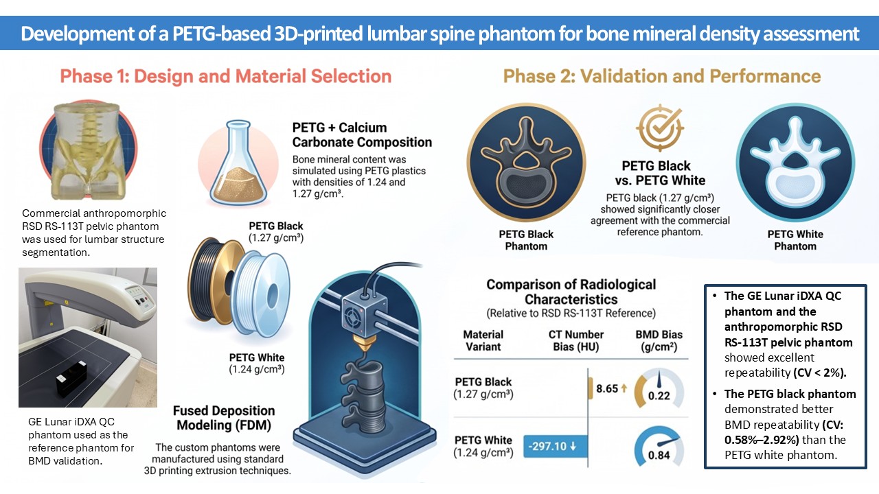
Materials and methods: A commercial RSD RS-113T pelvic phantom was scanned using CT and DXA to obtain reference CT numbers and BMD values. Lumbar spine structures were segmented from CT images to design a 3D-printed lumbar spine phantom using fused deposition modeling (FDM). Polyethylene terephthalate glycol (PETG; densities 1.24 and 1.27 gm/cm³), combined with calcium carbonate powder, was used to simulate bone mineral content. CT number and BMD measurements of the developed phantoms were compared with those of the RSD RS-113T pelvic phantom using Bland-Altman agreement analysis. BMD repeatability of the developed
phantoms was subsequently validated using the GE Lunar iDXA QC phantom.
Results: The PETG black phantom (1.27 gm/cm³) showed closer agreement with the RSD RS-113T pelvic phantom than the PETG white phantom (1.24 gm/cm³), with smaller Bland–Altman mean biases for CT number (8.65 vs -297.10 HU) and BMD (0.22 vs 0.84 gm/cm²). The RSD RS-113T pelvic phantom exhibited BMD CV values of less than 2%. The PETG black phantom also demonstrated better BMD repeatability (CV: 0.58%-2.92%) than the PETG white phantom, although some measurements exceeded the 2% precision criterion.
Conclusion: The PETG black 3D-printed lumbar spine phantom demonstrated superior BMD repeatability compared with the PETG white phantom. It also showed good agreement with the RSD RS-113T pelvic phantom and satisfactory BMD performance when validated using the GE Lunar iDXA QC phantom.
Article Details

This work is licensed under a Creative Commons Attribution-NonCommercial-NoDerivatives 4.0 International License.
Personal views expressed by the contributors in their articles are not necessarily those of the Journal of Associated Medical Sciences, Faculty of Associated Medical Sciences, Chiang Mai University.
References
Filippou V, Tsoumpas C. Recent advances on the development of phantoms using 3D printing for imaging with CT, MRI, PET, SPECT, and ultrasound. Med Phys. 2018; 45(9): e740-60. doi: 10.1002/mp.13058.
Okkalidis F, Chatzigeorgiou C, Okkalidis N, Dukov N, Milev M, Bliznakov Z, et al. Characterization of commercial and custom-made printing filament materials for computed tomography imaging of radiological phantoms. Technologies. 2024; 12(8): 139. doi: 10.3390/technologies12080139.
Pullen MW, Pooley RA, Kofler JM, Valero-Moreno F, Ramos-Fresnedo A, Domingo RA, et al. A radiographic analysis of common 3D print materials and assessment of their fidelity within vertebral models. Ann 3D Print Med. 2022; 8: 100080. doi: 10.1016/j.stlm.2022.100080.
Kim SY, Park JW, Park J, Yea JW, Oh SA. Fabrication of 3D printed head phantom using plaster mixed with polylactic acid powder for patient-specific QA in intensity-modulated radiotherapy. Sci Rep. 2022; 12: 17500. doi: 10.1038/s41598-022-22520-6.
Hatamikia S, Kronreif G, Unger A, Oberoi G, Jaksa L, Unger E, et al. 3D printed patient-specific thorax phantom with realistic heterogenous bone radiopacity using filament printer technology. Z Med Phys. 2022; 32(4): 438-52. doi: 10.1016/j.zemedi.2022.02.001.
Cochran BJ, Trajanovska S, Sunn N, Rye KA, Ryder WJ, editors. Development of 3D printing filaments for bespoke X-Ray phantoms. In: IEEE Nuclear Science Symposium and Medical Imaging Conference Proceedings (NSS/MIC); 2018 Nov 10-17; Sydney, NSW, Australia. IEEE; 2018.
Kozee M, Weygand J, Andreozzi JM, Hunt D, Perez BA, Graham JA, et al. Methodology for computed tomography characterization of commercially available 3D printing materials for use in radiology/radiation oncology. J Appl Clin Med Phys. 2023; 24(6): e13999. doi: 10.1002/acm2.13999.
Sangondimath G, Sen RK, T FR. DEXA and imaging in osteoporosis. Indian J Orthop. 2023; 57(Suppl 1): 82-93. doi: 10.1007/s43465-023-01059-2.
Slart RHJA, Punda M, Ali DS, Bazzocchi A, Bock O, Camacho P, et al. Updated practice guideline for dual-energy X-ray absorptiometry (DXA). Eur J Nucl Med Mol Imaging. 2024; 52: 539-63. doi: 10.1007/s00259-024-06912-6.
Frimeth J, Galiano E, Webster D. Some physical and clinical factors influencing the measurement of precision error, least significant change, and bone mineral density in dual-energy x-ray absorptiometry. J Clin Densitom. 2010; 13(1): 29-35. doi: 10.1016/j.jocd.2009.09.004.
Lewiecki EM, Binkley N, Morgan SL, Shuhart CR, Camargos BM, Carey JJ, et al. Best practices for dual-energy x-ray absorptiometry measurement and reporting: international society for clinical densitometry guidance. J Clin Densitom. 2016; 19(2): 127-40. doi: 10.1016/j.jocd.2016.03.003.
Kim HS, Yang SO. Quality control of DXA system and precision test of radio-technologists. J Bone Metab. 2014; 21(1): 2-7. doi: 10.11005/jbm.2014.21.1.2.
Mansour Z, Mokhtar A, Sarhan A, Ahmed MT, El-Diasty T. Quality control of CT image using American College of Radiology (ACR) phantom. Egypt J Radiol Nucl Med. 2016; 47(4): 1665-71. doi: 10.1016/j.ejrnm.2016.08.016.
Li Y, Jiang Y, Liu H, Yu X, Chen S, Ma D, et al. A phantom study comparing low-dose CT physical image quality from five different CT scanners. Quant Imaging Med Surg. 2022; 12(1): 766-80. doi: 10.21037/qims-21-245.
Hofmann T, Buschmann M, Homolka P. X-ray attenuation properties of additive manufacturing and 3d printing materials for mimicking tissues in radiographic phantoms measured by CT from 70 to 140 kV: 2025 update. Biomimetics (Basel). 2026; 11(3): 202. doi: 10.3390/biomimetics11030202.
Ahmed AM, Buschmann M, Breyer L, Kuntner C, Homolka P. Tailoring the mass density of 3D printing materials for accurate X-ray imaging simulation by controlled underfilling for radiographic phantoms. Polymers. 2024; 16(8): 1116. doi: 10.3390/polym16081116.
Jusufbegović M, Pandžić A, Šehić A, Jašić R, Julardžija F, Vegar-Zubović S, et al. Computed tomography tissue equivalence of 3D printing materials. Radiography (Lond). 2022; 28(3): 788-92. doi: 10.1016/j.radi.2022.02.008.
Mei K, Pasyar P, Geagan M, Liu LP, Shapira N, Gang GJ, et al. Design and fabrication of 3D-printed patient-specific soft tissue and bone phantoms for CT imaging. Sci Rep. 2023; 13(1): 17495. doi: 10.1038/s41598-023-44602-9.
Petraikin AV, Mikhailova AM, Kudryavtsev ND, Cherkasskaya MV, Yastrebova VO, Omelyanskaya OV, et al. Development of L1 vertebral anthropomorphic model for densitometric phantom improvement. Sovrem Tekhnologii Med. 2025; 17(4): 52-7. doi: 10.17691/stm2025.17.4.05 (in Russian).
Badiuk SR, Sasaki DK, Rickey DW. An anthropomorphic maxillofacial phantom using 3-dimensional printing, polyurethane rubber and epoxy resin for dental imaging and dosimetry. Dentomaxillofac Radiol. 2022; 51(1): 20200323. doi: 10.1259/dmfr.20200323.
