
The influence of 10 kVp and 15% rule applications on patient dose and image quality in extremities radiography: A phantom study
Article Sidebar
Main Article Content
Abstract
Background: Optimizing image quality and radiation dose is crucial in general radiography, adhering to the As Low As Reasonably Achievable (ALARA) principle.
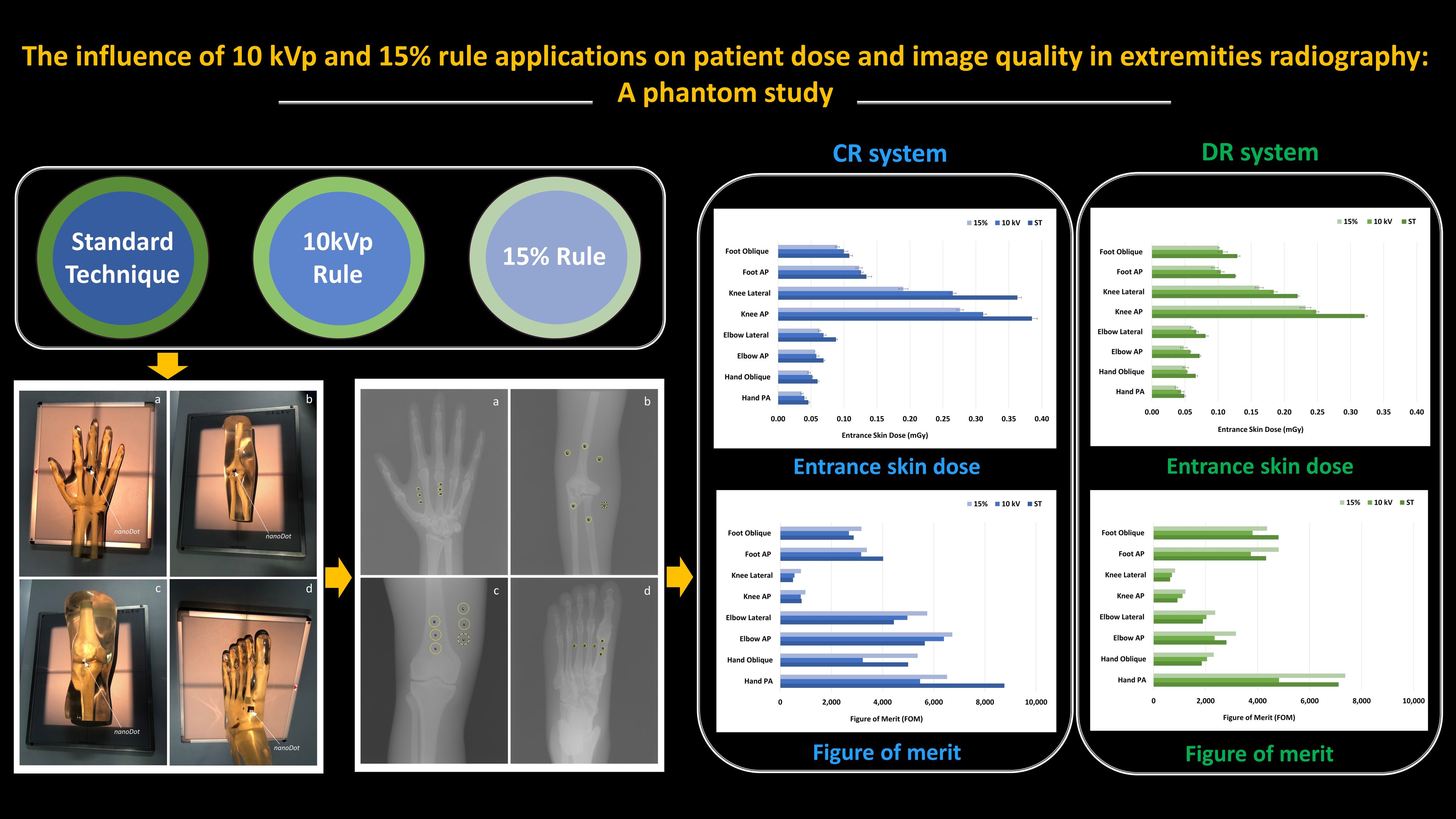
Objective: This study aimed to evaluate the impact of applying the 10 kVp and 15% rules on image quality and patient dose in extremity X-ray imaging using both computed radiography (CR) and digital radiography (DR) systems.
Materials and methods: X-ray imaging of hand, elbow, knee, and foot phantoms was performed using three different exposure techniques on both CR and DR systems. These techniques included the standard technique (ST) based on the established guidelines of the imaging systems, increased 10 kVp with a 50% mAs reduction from ST (10 kVp rule), and increased 15% kVp with a 50% mAs reduction from ST (15% rule). The entrance skin dose (ESD) was measured using nanoDot™placed on the phantom’s surface. The physical image qualities in contrast-to-noise ratio (CNR) and figure of merit (FOM) were utilized to assess the balance between image quality and radiation doses.
Results: The ESD was reduced by an average of -16% and -25% when applying the 10 kVp and 15% rules for all extremity imaging. This reduction decreased image CNR by -18% and -12%, respectively. There was no significant difference in CNR between the 15% and 10 kVp rule techniques for all extremity examinations in both CR and DR systems (p>0.05). Meanwhile, the exposure and deviation indexes remained within the established guidelines for CR and DR systems. However, the FOM values tended to be greater with the 15% rule technique than other techniques.
Conclusion: The ESD reduction was observed when applying the 10 kVp and 15% rules for all extremity imaging, both in CR and DR systems, with a slight degradation in image quality. The 15% rule represents the best option for optimization of image quality and patient dose based on the FOM results.
Article Details

This work is licensed under a Creative Commons Attribution-NonCommercial-NoDerivatives 4.0 International License.
Personal views expressed by the contributors in their articles are not necessarily those of the Journal of Associated Medical Sciences, Faculty of Associated Medical Sciences, Chiang Mai University.
References
Mettle r FA, Mahesh M, Bhargavan-Chatfield M, Chambers CE, Elee JG, Frush DP, et al. Patient exposure from radiologic and nuclear medicine procedures in the United States: Procedure volume and effective dose for the period 2006-2016. Radiology. 2020; 295(2): 418-27. doi: 10.1148/radiol.221263.
Lewis S, Pieterse T, Lawrence H. Retrospective evaluation of exposure indicators: a pilot study of exposure technique in digital radiography. J Med Radiat Sci. 2019; 66(1): 38-43. doi: 10.1002/jmrs.317.
Gibson DJ, Davidson RA. Exposure creep in computed radiography. A longitudinal study. Acad Radiol. 2012; 19(4): 458-62. doi: 10.1016/j.acra.2011.12.003.
Compagnone G, Baleni CM, Pagan L, Calzolaio FL, Barozzi L, Bergamini C. Comparison of radiation doses to patients undergoing standard radiographic examinations with conventional scree-film radiography, computed radiography and direct digital radiography. Br J Radiol. 2006; 79(947): 899-904. doi: 10.1259/bjr/57138583.
Seibert JA, Morin RL. The standardized exposure index for digital radiography: An opportunity for optimization of radiation dose to the pediatric population. Pediatr Radiol. 2011; 41(5): 573-81. doi: 10.1007/s00247-010-1954-6.
Ching W, Robinson J, Mcentee M. Patient-based radiographic exposure factor selection: A systematic review. J Med Radiat Sci. 2014; 61: 176-90. doi: 10.1002/jmrs.66.
Ching W, Robinson JW. DigiBit: A system for adjusting radiographic exposure factors in the digital era. Radio Technol. 2015; 86(6): 614-22. PMID: 26199434.
Martin CJ, Darragh CL, McKenzie GA, Bayliss AP. Implementation of a programme for reduction of radiographic doses and results achieved through increases in tube potential. Br J Radiol. 1993; 66(783): 228-33. doi: 10.1259/0007-1285-66-783-228.
Ramanaidu S, Sta Maria RB, Ng KH, George J, Kumar G. Evaluation of radiation dose and image quality following changes to tube potential (kVp) in conventional paediatric chest radiography. Biomed Imaging Interv J. 2006; 2(3): e35. doi: 10.2349/biij.2.3.e35
Coffey H, Chanopensiri V, Ly B, Nguyen D. Comparing 10 kVp and 15% Rules in Extremity Radiography. Radiol Technol. 2020; 91(6): 516-24. PMID: 32606229.
Brindhaban A, Al Khalifah K, Al Wathiqi G, Al Ostath H. Effect of x-ray tube potential on image quality and patient dose for lumbar spine computed radiography examinations. Australas Phys Eng Sci Med. 2005; 28(4): 216-22. doi: 10.1007/BF03178721.
Peacock NE, Steward AL, Riley PJ. An evaluation of the effect of tube potential on clinical image quality using direct digital detectors for pelvis and lumbar spine radiographs. J Med Radiat Sci. 2020; 67(4): 260-68. doi: 10.1002/jmrs.403.
Wenman A, Lockwood P. Comparing the standard knee X-ray exposure factor, 10 kV rule, and modified 10 kV rule techniques in digital radiography to reduce patient radiation dose without loss of image quality. Radiography. 2024; 30(2): 574-81. doi: 10.1016/j.radi.2024.01.013.
Moore QT, Don S, Goske MJ, Strauss KJ, Cohen M, Herrmann T, et al. Image gently: using exposure indicators to improve pediatric digital radiography. Radiol Technol. 2012; 84(1): 93-9. PMID: 22988269.
Vieworks. VXvue Operation Manual [Internet]. [cited 2024 Mar 21]. Available from: https://wosc.info/wp-content/uploads/EquipmentManuals/VXVUE/VXVUE-Operation-Manual.V86b21_EN-Manual.pdf
Freitas MB, Pimentel RB, Braga LF, Salido FSA, Neves RFCA, Medeiros RB. Patient dose optimization for computed radiography using physical and observerbased measurements as image quality metrics. Radiat Phys Chem. 2020; 172: 108768. doi: 10.1016/j.radphyschem.2020.108768.
Nocetti D, Villalobos K, Marín N, Monardes M, Tapia B, Toledo MI, et al. Radiation dose reduction and image quality evaluation for lateral lumbar spine projection. Heliyon. 2023; 9(9): e19509. doi: 10.1016/j.heliyon.2023.e19509.
