
Breast cancer characterization using region-based convolutional neural network with screening and diagnostic mammogram
Article Sidebar
Main Article Content
Abstract
Background: Detection and classification of microcalcifications in breast tissues is crucial for early breast cancer diagnosis and long-term treatment.
Objective: This paper aims to propose a robust model capable of detection and classification of breast cancer calcifications in digital mammogram images using Deep Convolutional Neural Networks (DCNN).
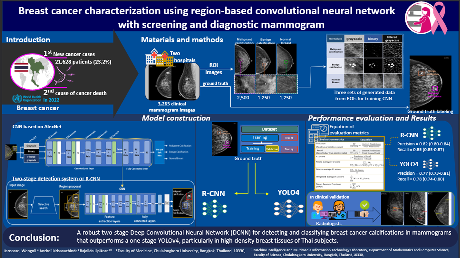
Materials and methods: An expert breast radiologist annotated the 3,265 clinical mammogram images to create a comprehensive ground truth dataset comprising 2,500 annotations for malignant and benign calcifications. This dataset was utilized to train our model, a two-stage detection system incorporating a Region-based Convolutional Neural Network (RCNN) with AlexNet and support vector machines to enhance the system’s robustness. The proposed model was compared to the one-stage detection, utilizing YOLOv4 combined with the Cross-Stage Partial Darknet53 (CSPDarknet53) architecture. A separate dataset of 504 mammogram images was explicitly set aside for model testing. The efficacy of the proposed model was evaluated based on key performance metrics, including precision, recall, F1 score, and mean average precision (mAP).
Results: The results showed that the proposed RCNN-2 model could automatically identify and categorize calcifications as malignant or benign, outperforming the YOLOv4 models. The RCNN-2’s overall effectiveness, as evaluated by precision, recall, F1 score, and mean average precision (mAP), achieved scores of 0.82, 0.85, 0.83, and 0.74, respectively.
Conclusion: The proposed RCNN-2 model demonstrates very effective detection and classification of calcification in mammogram images, especially in high-dense breast images. The performance of the proposed model was compared to that of YOLOv4, and it can be concluded that the proposed RCNN model yields outstanding performance. The model can be a helpful tool for radiologists.
Article Details

This work is licensed under a Creative Commons Attribution-NonCommercial-NoDerivatives 4.0 International License.
Personal views expressed by the contributors in their articles are not necessarily those of the Journal of Associated Medical Sciences, Faculty of Associated Medical Sciences, Chiang Mai University.
References
Ferlay J, Ervik M, Lam F, Laversanne M, Colombet M, Mery L, et al. Global Cancer Observatory: Cancer Today.
Lyon, France: International Agency for Research on Cancer. 2024. https://gco.iarc.fr/today [accessed 1 February 2024].
Padia K, Douglas TS, Cairncross LL, Baasch RV, Vaughan CL. Detecting Breast Cancer with a DualModality Device. Diagnostics. 2017; 7(1): 17. doi: 10.3390/diagnostics7010017
Haus AG, Yaffe MJ. Screen-Film and Digital Mammography: Image Quality and Radiation Dose Considerations. Radiol Clin North Am. 2000; 38(4): 871-98. doi: 10.1016/s0033-8389(05)70207-4
Sechopoulos I. A review of breast tomosynthesis. Part I. The image acquisition process. Med Phys. 2013; 40(1): 014301. doi: 10.1118/1.4770279
International Atomic Energy Agency, Quality Assurance Programme for Digital Mammography, IAEA Human Health Series No. 17, IAEA, Vienna, 2011.
Yilmaz R, Aydiner A, Igci A, Soran A. Breast Imaging: Breast Cancer A Guide to Clinical Practice. 1st ed. Springer Nature, Switzerland, AG, 2019.
American College of Radiology, Mammography saves lives. https://www.acr.org/Practice-ManagementQuality-Informatics/Practice-Toolkit/Patient-Resources/Mammography-Saves-Lives. Accessed on February 20, 2023.
Monticciolo DL, Newell MS, Hendrick RE, Helvie MA, Moy L, Monsees B, et al. Breast Cancer Screening for Average-Risk Women: Recommendations From the ACR Commission on Breast Imaging. J Am Coll Radiol. 2017; 14: 1137-43. doi: 10.1016/j.jacr.2017.06.001
Nyante SJ, Lee SS, Benefield T, Hoots TN, Henderson LM. The association between mammographic calcifications and breast cancer prognostic factors in a population-based registry cohort. Cancer. 2017; 123: 219-27. doi: 10.1002/cncr.30281
Logullo AF, Prigenzi KCK, Nimir CCBA, Franco AFV, Campos MSDA. Breast microcalcifications: Past, present and future (Review). Mol Clin Oncol. 2022; 16(4): 81. doi: 10.3892/mco.2022.2514
Cai H, Huang Q, Rong W, Song Y, Li J, Wang J. et al. Breast Microcalcification Diagnosis Using Deep Convolutional Neural Network from Digital Mammograms. Comput Math Methods Med. 2019; 1-10. doi: 10.1155/2019/2717454
Chen Z, Strange H, Oliver A, Denton ER, Boggis C, Zwiggelaar R, et al. Topological Modeling and Classification of Mammographic Microcalcification Clusters. IEEE Trans Biomed Eng. 2015; 62: 1203-14. doi: 10.1109/TBME.2014.2385102
Singh N, Marak J, Joshi P, Singh DK. Morphological and distribution pattern of calcifications on full field digital mammography versus digital breast tomosynthesis and comparison of diagnostic abilities of the two modalities: A retrospective study. J Clin of Diagn Res. 2023; 17(3): TC36-TC41. doi: 10.7860/JCDR/2023/55632.17675
Mann RM, Athanasiou A, Baltzer PAT, et al. Breast cancer screening in women with extremely dense breasts recommendations of the European Society of Breast Imaging (EUSOBI). Eur Radiol. 2022; 32(6): 4036-45. doi: 10.1007/s00330-022-08617-6
Edmonds CE, O’Brien SR, Conant EF,et al. Mammographic Breast Density: Current Assessment Methods, Clinical Implications, and Future Directions. Seminars in Ultrasound, CT, and MRI. Epub. 2022; 44: 35-45. doi: 10.1053/j.sult.2022.11.001
Sickles EA, D’Orsi CJ, Bassett LW. American College of Radiology. ACR BI-RADS® Atlas, 5th ed. Reston, VA, USA, 2013.
Gordon PB. The Impact of Dense Breasts on the Stage of Breast Cancer at Diagnosis: A Review and Options for Supplemental Screening. Curr Oncol. 2022; 17; 29(5): 3595-636. doi: 10.3390/curroncol29050291
Bodewes FTH, van Asselt AA, Dorrius MD, Greuter MJW, de Bock GH. Mammographic breast density and the risk of breast cancer: A systematic review and meta-analysis. Breast. 2022; 66: 62-8. doi: 10.1016/j.breast.2022.09.007
Sarvamangala DR, Kulkarni RV. Convolutional neural networks in medical image understanding: a survey. Evol Intell. 2022; 15(1): 1-22. doi: 10.1007/s12065-020-00540-3
Yamashita R, Nishio M, Do RKG, Togashi K. Convolutional neural networks: an overview and application in radiology. Insights Imaging. 2018; 9; 611-29. doi: 10.1007/s13244-018-0639-9
Ahmed SF, Alam MSB, Hassan M, Rozbu MR, Ishtiak T, Rafa N, et al. Deep learning modelling techniques: current progress, applications, advantages, and challenges. Artif Intell Rev. 2023; 56: 13521-617. doi: 10.1007/s10462-023-10466-8
Aphinives C, Aphinives P, Nawapan S. Artificial Intelligence Development for Detecting Microcalcification within Mammography. J Med Assoc Thai. 2021; 104(4): 560-4. doi: 10.1002/jmat.2021.104.560
Intasam A, Promworn Y, Thanasitthichai S, Piyawattanametha W. A comparative study of convolutional neural networks for mammogram diagnosis. Proceedings of 14th the Biomedical Engineering International Conference (BMEiCON-2022); 2022 Nov 10-13; Songkhla, Thailand. p.1-4. doi: 10.1109/BMEiCON56653.202210012074. Available from: http://ieeexplore. ieee.org/document/10012074.
Labcharoenwongs P, Vonganansup S, Chunhapran O, Noolek D, Yampaka T. An Automatic Breast Tumor Detection and Classification including Automatic Tumor Volume Estimation Using Deep Learning Technique. Asian Pac J Cancer Prev. 2023; 24(3): 1081-8. doi: 10.31557/APJCP.2023.24.3.1081
Lehman CD, Yala A, Schuster T, Dontchos B, Bahl M, Swanson K, et al. Mammographic Breast Density Assessment Using Deep Learning: Clinical Implementation. Radiol. 2019; 290(1): 52-8. doi.org/10.1148/radiol.2018180694
Sabani A, Landsmann A, Hejduk P, Schmidt C, Marcon M, Borkowski K, et al. BI-RADS-Based Classification of Mammographic Soft Tissue Opacities Using a Deep Convolutional Neural Network. Diagnostics. 2022; 12(7): 1564. doi: 10.3390/diagnostics12071564
Zitnick CL, Dollár P. Edge Boxes: Locating Object Proposals from Edges. European Conference on Computer Vision. 2014: Part V, LNCS 8693 p.391-405.
