
Comparative study of Immunohistochemistry (IHC) staining of Anatomical Pathology Saraburi Hospital with staining by automatic machine
Abstract
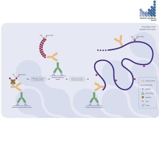
Immunohistochemistry (IHC) is an anatomical pathology laboratory technique for staining tissue samples using a special method that utilizes the antigen-antibody reaction 1 to detect antigens on the tissue and produce a visible color signal under a microscope2. IHC is commonly used in disease diagnosis, disease sign detection, and treatment response prediction.1 It is widely used in anatomical pathology laboratories, and the Surgical Pathology Laboratory, Anatomical Pathology Department, Saraburi Hospital, is one such unit that has adopted the IHC method to aid in pathological diagnosis. Its high specificity allows pathologists to differentiate between various cell types, contributing to more accurate disease diagnosis. Currently, many modern staining techniques and automated staining machines are available from various companies. Therefore, this research aims to compare the traditional staining method used by the anatomical pathology laboratory at Saraburi Hospital with the automated staining method to determine the efficiency and significance of IHC staining, and to establish a quality standard for operational practices. The experiment compared IHC staining with a total of 30 antibodies and analyzed the data using the Independent T-Test. The experiment was divided into two groups: Group 1 used manual immunohistochemical staining (Protocol A), and Group 2 used an automated immunohistochemical staining machine (Protocol B). Each group used 30 antibodies and the same dilution. Three subgroups were considered: positive control, unknown, and negative control. The results showed no significant difference between the two positive control groups (Sig.<0.170). The unknown group showed a significant difference (Sig.<0.012), and the negative control group showed no significant difference. This concludes that the IHC staining method used by the Anatomical Pathology Department at Saraburi Hospital is efficient and meets the standards of automated IHC staining widely used in both public and private anatomical pathology departments.
References
Bancroft JD. Theory and practice of histological techniques. 3rd ed.Edinburg :Churchill Livingstone , 1990.
Gustavson MD, Bourke-Martin B, Reilly D, et al. Standardization of HER2 immunohistochemistry in breast cancer by automated quantitative analysis. Arch Pathol Lab Med. 2009;133:1413–9. doi: 10.5858/133.9.1413.
Kim SW, Roh J, Park CS. Immunohistochemistry for Pathologists: Protocols, Pitfalls, and Tips. J Pathol Transl Med. 2016 Nov;50(6):411-418. doi: 10.4132/jptm.2016.08.08. Epub 2016 Oct 13. PMID: 27809448; PMCID: PMC5122731.
Tzankov A, Zlobec I, Went P, Robl H, Hoeller S, Dirnhofer S. Prognostic immunophenotypic biomarker studies in diffuse large B cell lymphoma with special emphasis on rational determination of cut-off scores. Leuk Lymphoma. 2010;51:199–212. doi: 10.3109/10428190903370338. [DOI] [PubMed] [Google Scholar]
Yaziji H, Barry T. Diagnostic Immunohistochemistry: what can go wrong? Adv Anat Pathol. 2006;13:238–46. doi: 10.1097/01.pap.0000213041.39070.2f. [DOI] [PubMed] [Google Scholar]
วิจัยทางการแพทย์/ธีระพร วุฒยวนิช ,นิมิต มรกต , กิตติกา กาญจนรัตนากร บรรณาธิการ. พิมพ์ครั้งที่ 2 . เชียงใหม่ : โครงการตำรา : คณะแพทยศาสตร์ มหาวิทยาลัยเชียงใหม่ , 2553
Catalog 2011.Product & Services. Dako.139.
https://www.celnovte.com/news/automated-vs-manual-ihc-stainers-time-cost-error-rates-in-high-throughput-labs/#:~:text=*%20Product.%20*%20Solution . เข้าถึงเมื่อวันที่ 21/12/2568
Cell MarqueTM Tissue Diagnostics Immunohistochemistry Reference Guide Vol.12
https://www.sciencedirect.com/science/article/abs/pii/S0009898198001466 เข้าถึงเมื่อวันที่ 24/12/2568
Đorđević M, Životić M, Radojević Škodrić S, Nešović Ostojić J, Marković Lipkovski J, Filipović J, Ćirović S, Kovačević S, Dunđerović D. Effects of Automation on Sustainability of Immunohistochemistry Laboratory. Healthcare (Basel). 2021 Jul 8;9(7):866. doi: 10.3390/healthcare9070866. PMID: 34356244; PMCID: PMC8304755.
https://pmc.ncbi.nlm.nih.gov/articles/PMC8304755/
Streitberg G.S., Angel L., Sikaris K.A., Bwititi P.T. Automation in clinical biochemistry: Core, peripheral, STAT, and specialist laboratories in Australia. J. Lab. Autom. 2012;17:387–394. doi: 10.1177/2211068212448865. [DOI] [PubMed] [Google Scholar]
Downloads
Published
How to Cite
Issue
Section
License
Copyright (c) 2026 Siraporn Ruangvech

This work is licensed under a Creative Commons Attribution-NonCommercial-NoDerivatives 4.0 International License.
Journal of TCI is licensed under a Creative Commons Attribution-NonCommercial-NoDerivatives 4.0 International (CC BY-NC-ND 4.0) licence, unless otherwise stated. Please read our Policies page for more information.


