
การศึกษาเปรียบเทียบทางกายวิภาคของหลอดเลือดดำคอข้างขวาและข้างซ้าย: เพื่อเป็นแนวทางในการใส่สายสวนหลอดเลือดดำใหญ่
Article Sidebar
Main Article Content
บทคัดย่อ
บทนำ: สายสวนหลอดเลือดดำส่วนกลางมีความจำเป็นสำหรับการฟอกไต หลอดเลือดดำคอ (internal jugular vein, IJV) เป็นตำแหน่งที่นิยมใช้ในการเข้าถึงหลอดเลือด และจำเป็นต้องทราบถึงความหลากหลายทางกายวิภาคของหลอดเลือดนี้เพื่อป้องกันการแทงพลาดเข้าหลอดเลือดแดงโดยอุบัติเหตุ การศึกษานี้มีวัตถุประสงค์เพื่ออธิบายความหลากหลายทางกายวิภาคของหลอดเลือดดำคอทั้งสองข้าง
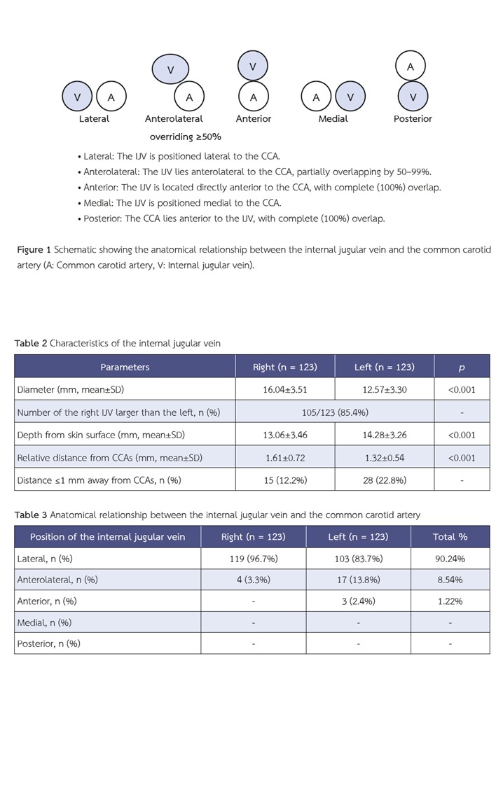
ระเบียบวิธีวิจัย: การศึกษานี้เป็นการศึกษาทบทวนข้อมูลผู้ป่วยย้อนหลังจำนวน 123 ราย ที่ได้รับการตรวจเอกซเรย์คอมพิวเตอร์บริเวณคอ ตั้งแต่วันที่ 1 มกราคม พ.ศ. 2558 ถึง 31 ธันวาคม พ.ศ. 2562 โดยบันทึกข้อมูลขนาดเส้นผ่านศูนย์กลางของหลอดเลือดดำคอทั้งสองข้าง ความลึกจากผิวหนัง ระยะห่างจากหลอดเลือดแดงคาโรติด และตำแหน่งของหลอดเลือดดำคอเทียบกับหลอดเลือดแดงคาโรติด จุดอ้างอิงสำหรับตำแหน่งของหลอดเลือดดำคอคือหลอดเลือดแดงคาโรติด โดยจำแนกตำแหน่งเป็น ด้านข้าง (lateral) ด้านหน้า (anterior) ด้านหน้าเฉียงข้าง (anterolateral) ด้านใน (medial) หรือด้านหลัง (posterior) ประเมินที่ระดับกระดูกอ่อนไครคอยด์
ผลการวิจัย: หลอดเลือดดำคอส่วนใหญ่ (90.24%) อยู่ทางด้านข้างของหลอดเลือดแดงคาโรติด โดยหลอดเลือดดำคอข้างซ้าย แสดงความหลากหลายของตำแหน่งมากกว่า หลอดเลือดดำคอข้างซ้ายมีการซ้อนทับกับหลอดเลือดแดงคาโรติดในตำแหน่งด้านหน้าเฉียงข้างบ่อยกว่าข้างขวา พบหลอดเลือดดำคอข้างซ้ายเท่านั้นที่อยู่ในตำแหน่งด้านหน้า หลอดเลือดดำคอข้างขวามีขนาดใหญ่กว่า (85.4%) โดยมีขนาดเส้นผ่านศูนย์กลางแตกต่างกันอย่างมีนัยสำคัญ (16.04±3.51 มม. เทียบกับ 12.57±3.30 มม., p <0.001) และ อยู่ตื้นกว่า (13.06±3.46 มม. เทียบกับ 14.28±3.26 มม., p <0.001)
สรุป: หลอดเลือดดำคอข้างขวาเหมาะสมสำหรับการเข้าถึงหลอดเลือดมากกว่าข้างซ้าย เนื่องจากมีความหลากหลายทางกายวิภาคน้อยกว่าและมีตำแหน่งที่เอื้ออำนวยต่อการแทงมากกว่า
Article Details

อนุญาตภายใต้เงื่อนไข Creative Commons Attribution-NonCommercial-NoDerivatives 4.0 International License.
บทความนี้ตีพิมพ์ภายไต้การอนุญาต CC BY-NC-ND 4.0 ซึ่งอนุญาตให้สามารถใช้บทความนี้พื่อวัตถุประสงค์ใดๆ ก็ตามที่ไม่ใช่เชิงพาณิชย์ โดยต้องมีการอ้างถึงที่มาของบทความอย่างครบถ้วน ใครก็ตามสามารถคัดลอกและแจกจ่ายทุกส่วนของบทความนี้โดยไม่ต้องขออนุญาตจากผู้ประพันธ์หรือสมาคมโรคไตแห่งประเทศไทย
เอกสารอ้างอิง
Diseases GBD, Injuries C. Global burden of 369 diseases and injuries in 204 countries and territories, 1990-2019: a systematic analysis for the Global Burden of Disease Study 2019. Lancet 2020;396(10258):1204–22. doi: 10.1016/S0140-6736(20)30925-9.
Kovesdy CP. Epidemiology of chronic kidney disease: an update 2022. Kidney Int Suppl (2011) 2022;12(1):7–11. doi: 10.1016/j.kisu.2021.11.003.
Kidney Disease: Improving Global Outcomes CKDWG. KDIGO 2024 Clinical Practice Guideline for the Evaluation and Management of Chronic Kidney Disease. Kidney Int 2024;105(4S):S117–S314. doi: 10.1016/j.kint.2023.10.018.
Lok CE, Huber TS, Lee T, Shenoy S, Yevzlin AS, Abreo K, et al. KDOQI Clinical Practice Guideline for Vascular Access: 2019 Update. Am J Kidney Dis 2020;75(4 Suppl 2):S1–S164. doi: 10.1053/j.ajkd.2019.12.001.
McGee DC, Gould MK. Preventing complications of central venous catheterization. N Engl J Med 2003;348(12):1123–33. doi: 10.1056/NEJMra011883.
Vats HS. Complications of catheters: tunneled and nontunneled. Adv Chronic Kidney Dis 2012;19(3):188–94. doi: 10.1053/j.ackd.2012.04.004.
Schillinger F, Schillinger D, Montagnac R, Milcent T. Post catheterisation vein stenosis in haemodialysis: comparative angiographic study of 50 subclavian and 50 internal jugular accesses. Nephrol Dial Transplant 1991;6(10):722–4. doi: 10.1093/ndt/6.10.722.
Bowdle A. Vascular complications of central venous catheter placement: evidence-based methods for prevention and treatment. J Cardiothorac Vasc Anesth 2014;28(2):358–68. doi: 10.1053/j.jvca.2013.02.027.
Safety Committee of Japanese Society of A. Practical guide for safe central venous catheterization and management 2017. J Anesth 2020;34(2):167–86. doi: 10.1007/s00540-019-02702-9.
Lim CL, Keshava SN, Lea M. Anatomical variations of the internal jugular veins and their relationship to the carotid arteries: a CT evaluation. Australas Radiol 2006;50(4):314–8. doi: 10.1111/j.1440-1673.2006.01589.x.
Maneenai N, Arjhansiri K. CT evaluation of anatomical variations of the internal jugular veins in Thai adults. Asian Biomedicine 2013;7(6):803–11. doi: 10.5372/1905-7415.0706.243.
Kornbau C, Lee KC, Hughes GD, Firstenberg MS. Central line complications. Int J Crit Illn Inj Sci 2015;5(3):170–8. doi: 10.4103/2229-5151.164940.
Jin Lee J, Sook Gwak M, Yang M, Soo Kim G. A new method of internal jugular vein catheterization using the cricoid cartilage and the external jugular vein as a landmark. Am J Emerg Med 2006;24(6):697–701. doi: 10.1016/j.ajem.2006.03.005.
Buch K, Groller R, Nadgir RN, Fujita A, Qureshi MM, Sakai O. Variability in the Cross-Sectional Area and Narrowing of the Internal Jugular Vein in Patients Without Multiple Sclerosis. AJR Am J Roentgenol 2016;206(5):1082–6. doi: 10.2214/AJR.15.14689.
Bannon MP, Heller SF, Rivera M. Anatomic considerations for central venous cannulation. Risk Manag Healthc Policy 2011;4:27–39. doi: 10.2147/RMHP.S10383.
Ayres A, van Tonder DJ, van Schoor A-N. Anatomical analysis of Sedillot’s triangle as a reliable landmark for insertion of central venous catheters in neonates using a central approach. Translational Research in Anatomy 2023;33:100264. doi: 10.1016/j.tria.2023.100264.
Troianos CA, Kuwik RJ, Pasqual JR, Lim AJ, Odasso DP. Internal jugular vein and carotid artery anatomic relation as determined by ultrasonography. Anesthesiology 1996;85(1):43–8. doi: 10.1097/00000542-199607000-00007.
Lin BS, Kong CW, Tarng DC, Huang TP, Tang GJ. Anatomical variation of the internal jugular vein and its impact on temporary haemodialysis vascular access: an ultrasonographic survey in uraemic patients. Nephrol Dial Transplant 1998;13(1):134–8. doi: 10.1093/ndt/13.1.134.
Saugel B, Scheeren TWL, Teboul JL. Ultrasound-guided central venous catheter placement: a structured review and recommendations for clinical practice. Crit Care 2017;21(1):225. doi: 10.1186/s13054-017-1814-y.
Sulek CA, Blas ML, Lobato EB. A randomized study of left versus right internal jugular vein cannulation in adults. J Clin Anesth 2000;12(2):142–5. doi: 10.1016/s0952-8180(00)00129-x.
Asouhidou I, Natsis K, Asteri T, Sountoulides P, Vlasis K, Tsikaras P. Anatomical variation of left internal jugular vein: clinical significance for an anaesthesiologist. Eur J Anaesthesiol 2008;25(4):314–8. doi: 10.1017/S0265021508003700.
Saiki K, Tsurumoto T, Okamoto K, Wakebe T. Relation between bilateral differences in internal jugular vein caliber and flow patterns of dural venous sinuses. Anat Sci Int 2013;88(3):141–50. doi: 10.1007/s12565-013-0176-z.
Kosnik N, Kowalski T, Lorenz L, Valacer M, Sakthi-Velavan S. Anatomical review of internal jugular vein cannulation. Folia Morphol (Warsz) 2024;83(1):1–19. doi: 10.5603/FM.a2023.0008.
Lorchirachoonkul T, Ti LK, Manohara S, Lye ST, Tan SA, Shen L, et al. Anatomical variations of the internal jugular vein: implications for successful cannulation and risk of carotid artery puncture. Singapore Med J 2012;53(5):325–8.
Patel AR, Patel AR, Singh S, Singh S, Khawaja I. Central Line Catheters and Associated Complications: A Review. Cureus 2019;11(5):e4717. doi: 10.7759/cureus.4717.
Kwon SS, Falk A, Mitty HA. Thoracic duct injury associated with left internal jugular vein catheterization: anatomic considerations. J Vasc Interv Radiol 2002;13(3):337–9. doi: 10.1016/s1051-0443(07)61730-8.
Merritt RL, Hachadorian ME, Michaels K, Zevallos E, Mhayamaguru KM, Closser Z, et al. The Effect of Head Rotation on the Relative Vascular Anatomy of the Neck: Implications for Central Venous Access. J Emerg Trauma Shock 2018;11(3):193–6. doi: 10.4103/JETS.JETS_5_18.
