The Brain’s Gendered Architecture: An MRI Study of Cortical and Subcortical Volume Differences
DOI:
https://doi.org/10.12982/BSCM.2026.44Keywords:
sex differences, cortical and subcortical volumes, MRI, brain morphometry, neuroanatomyAbstract
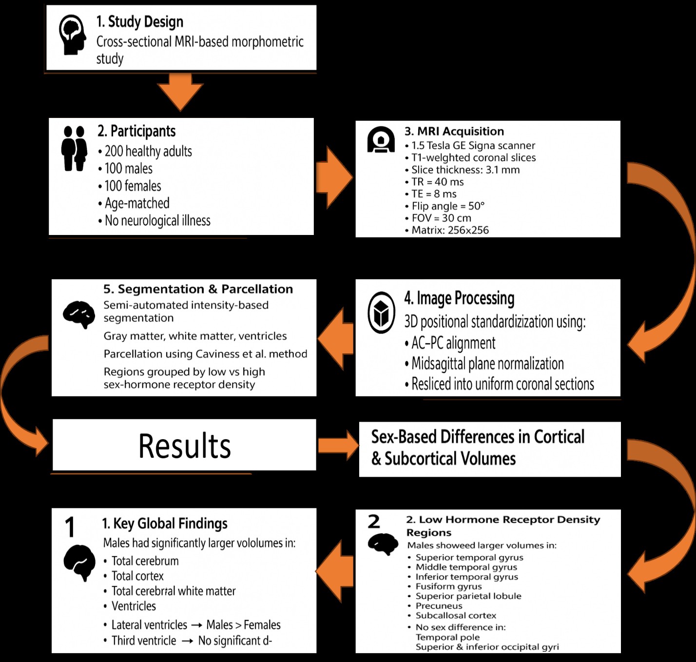
Objective For the advancement of neuroscience and therapeutic applications, it is crucial to understand how cerebral and subcortical volumes change across sexes. This study utilized magnetic resonance imaging (MRI) to investigate volumetric variations in various brain areas across different sexes.
Methods A cross-sectional study was conducted on 200 healthy participants (100 males and 100 females). High-resolution MRI scans were acquired using a 1.5T General Electric Signa scanner, and cortical segmentation was performed using a standardized semi-automated method. Statistical analyses were conducted using IBM SPSS, with independent sample t-tests employed to compare volumetric differences between sexes. Effect sizes were computed using Cohen’s d, and multiple comparisons were adjusted via the Bonferroni method.
Results There were considerably greater volumes of total cerebral white matter (p < 0.001, d = -1.1), total cortex (p < 0.001, d = -0.635), and total cerebrum (p < 0.001, d = -1.352) in males. Males also had considerably bigger ventricular structures, such as the lateral (p < 0.001, d = -0.71) and third (p < 0.001, d = -1.229) ventricles, although there was no significant difference in the fourth ventricle (p = 0.171). Males had substantially greater volumes in the inferior temporal gyrus (p < 0.001, d = -0.58), middle temporal gyrus (p < 0.001, d = -0.55), and superior temporal gyrus (p < 0.001, d = -0.73) among cortical areas with low-density sex hormone receptors. Significant differences were seen in the occipital pole (p < 0.001, d = -1.342), thalamus (p < 0.001, d = -1.282), and postcentral gyrus (p < 0.001, d = -1.536) in areas with high-density sex hormone receptors.
Conclusions This study confirms significant sex-based differences in brain volume, particularly in cortical and subcortical structures. These findings highlight the role of sex hormones in shaping neuroanatomy and provide insights for future research on sex-specific neurological and psychiatric conditions.
References
Allen JS, Damasio H, Grabowski TJ, Bruss J, Zhang W. Sexual dimorphism and asymmetries in the gray–white composition of the human cerebrum. Neuroimage. 2003;18:880–94.
Ruigrok ANV, Salimi-Khorshidi G, Lai M-C, Baron-Cohen S, Lombardo M V, Tait RJ, et al. A meta-analysis of sex differences in human brain structure. Neurosci Biobehav Rev. 2014;39:34–50.
Luders E, Narr KL, Thompson PM, Rex DE, Woods RP, DeLuca H, et al. Gender effects on cortical thickness and the influence of scaling. Hum Brain Mapp. 2006; 27:314–24.
Goldstein JM, Seidman LJ, Horton NJ, Makris N, Kennedy DN, Caviness Jr VS, et al. Normal sexual dimorphism of the adult human brain assessed by in vivo magnetic resonance imaging. Cereb Cortex. 2001;11: 490–7.
Ramzanpour M, Jafari B, Smith J, Allen J, Hajiaghamemar M. Comprehensive study of sex-based anatomical variations of human brain and development of sex-specific brain templates. Brain Multiphysics. 2023;4:100077.
Parsaei M, Sanjari Moghaddam H, Aarabi MH. Sex differences in brain structures throughout the lifetime. Aging Brain. 2023;4:100098. PubMed PMID: 37809276
Del Mauro G, Del Maschio N, Sulpizio S, Fedeli D, Perani D, Abutalebi J. Investigating sexual dimorphism in human brain structure by combining multiple indexes of brain morphology and source-based morphometry. Brain Struct Funct. 2022;227:11–21.
Williams CM, Peyre H, Toro R, Ramus F. Sex differences in the brain are not reduced to differences in body size. Neurosci Biobehav Rev. 2021;130:509–11.
Sanchis-Segura C, Aguirre N, Cruz-Gómez ÁJ, Félix S, Forn C. Beyond “sex prediction”: Estimating and interpreting multivariate sex differences and similarities in the brain. Neuroimage. 2022;257:119343.
Cosgrove KP, Mazure CM, Staley JK. Evolving Knowledge of Sex Differences in Brain Structure, Function, and Chemistry. Biol Psychiatry. 2007;62:847–55.
Filipek PA, Richelme C, Kennedy DN, Caviness VS. The Young Adult Human Brain: An MRI-based Morphometric Analysis. Cereb Cortex. 1994;4:344–60.
Caviness VS, Meyer J, Makris N, Kennedy DN. MRI-based topographic parcellation of human neocortex: an anatomically specified method with estimate of reliability. J Cogn Neurosci. 1996;8:566–87.
Goldstein JM, Goodman JM, Seidman LJ, Kennedy DN, Makris N, Lee H, et al. Cortical abnormalities in schizophrenia identified by structural magnetic resonance imaging. Arch Gen Psychiatry. 1999;56:537–47.
Rademacher J, Galaburda AM, Kennedy DN, Filipek PA, Caviness VS. Human Cerebral Cortex: Localization, Parcellation, and Morphometry with Magnetic Resonance Imaging. J Cogn Neurosci. 1992;4:352–74.
Seidman LJ, Faraone SV, Goldstein JM, Goodman JM, Kremen WS, Toomey R, et al. Thalamic and amygdala–hippocampal volume reductions in first-degree relatives of patients with schizophrenia: an MRI-based morphometric analysis. Biol Psychiatry. 1999;46:941–54.
Ingalhalikar M, Smith A, Parker D, Satterthwaite TD, Elliott MA, Ruparel K, et al. Sex differences in the structural connectome of the human brain. Proc Natl Acad Sci U S A. 2014;111:823–8.
Cowell PE, Sluming VA, Wilkinson ID, Cezayirli E, Romanowski CAJ, Webb JA, et al. Effects of sex and age on regional prefrontal brain volume in two human cohorts. Eur J Neurosci. 2007;25:307–18.
Soysal H, Acer N, Özdemir M, Eraslan Ö. Volumetric measurements of the subcortical structures of healthy adult brains in the Turkish population. Folia Morphol (Warsz). 2022;81:294–306.
Adeli E, Zhao Q, Zahr NM, Goldstone A, Pfefferbaum A, Sullivan EV, et al. Deep learning identifies morphological determinants of sex differences in the pre-adolescent brain. Neuroimage. 2020;223:117293. PubMed PMID: 32841716
Mondal M, Das R, Mukherjee A. Variations of normal ventricular indices depending on age and sex by computed tomography. Int J Sci Res. 2021;10:52-5.
Backhausen LL, Fröhner JH, Lemaître H, Artiges E, Martinot M-LP, Herting MM, et al. Adolescent to young adult longitudinal development of subcortical volumes in two European sites with four waves. Hum Brain Mapp. 2024;45:e26574. PubMed PMID: 38401132
Giedd JN, Raznahan A, Mills KL, Lenroot RK. Review: magnetic resonance imaging of male/female differences in human adolescent brain anatomy. Biol Sex Differ. 2012;3:19. PubMed PMID: 22908911
Nuñez JL, Huppenbauer CB, McAbee MD, Juraska JM, DonCarlos LL. Androgen receptor expression in the developing male and female rat visual and prefrontal cortex. J Neurobiol. 2003;56:293–302.
Raznahan A, Lee Y, Stidd R, Long R, Greenstein D, Clasen L, et al. Longitudinally mapping the influence of sex and androgen signaling on the dynamics of human cortical maturation in adolescence. Proc Natl Acad Sci U S A. 2010;107:16988–93.
Long M, Ostertag C, Reynolds JE, Zheng J, Landman B, Huo Y, et al. Few sex differences in regional gray matter volume growth trajectories across early childhood. Imaging Neurosci. 2024;2:1–26.
Küchenhoff S, Bayrak Ş, Zsido RG, Saberi A, Bernhardt BC, Weis S, et al. Relating sex-bias in human cortical and hippocampal microstructure to sex hormones. Nat Commun. 2024;15:7279. PubMed PMID: 39179555
Cara AL, Henson EL, Beekly BG, Elias CF. Distribution of androgen receptor mRNA in the prepubertal male and female mouse brain. J Neuroendocrinol 2021;33:e13063. PubMed PMID: 34866263
Campbell CE, Mezher AF, Tyszka JM, Nagel BJ, Eckel SP, Herting MM. Associations between testosterone, estradiol, and androgen receptor genotype with amygdala subregions in adolescents. Psychoneuroendocrinology 2022;137:105604. PubMed PMID: 34971856
Peper JS, Brouwer RM, Schnack HG, van Baal GC, van Leeuwen M, van den Berg SM, et al. Sex steroids and brain structure in pubertal boys and girls. Psychoneuroendocrinology. 2009;34:332–42.
Tan GC-Y, Chu C, Lee YT, Tan CCK, Ashburner J, Wood NW, et al. The influence of microsatellite polymorphisms in sex steroid receptor genes ESR1, ESR2 and AR on sex differences in brain structure. Neuroimage. 2020;221:117087. PubMed PMID: 32593802
van Eijk L, Hansell NK, Strike LT, Couvy-Duchesne B, de Zubicaray GI, Thompson PM, et al. Region-specific sex differences in the hippocampus. Neuroimage. 2020;215:116781. PubMed PMID: 32278894
López-Ojeda W, Hurley RA. Sexual Dimorphism in Brain Development: Influence on Affective Disorders. J Neuropsychiatry Clin Neurosci. 2021;33:A485-9.