Repeatability and Reproducibility of an Automated Image-Processing Software for Quantitative Conjunctival Hyperemia Analysis: A Pilot Study
DOI:
https://doi.org/10.12982/BSCM.2026.38Keywords:
repeatability, reproducibility, conjunctival hyperemia, automated, softwareAbstract
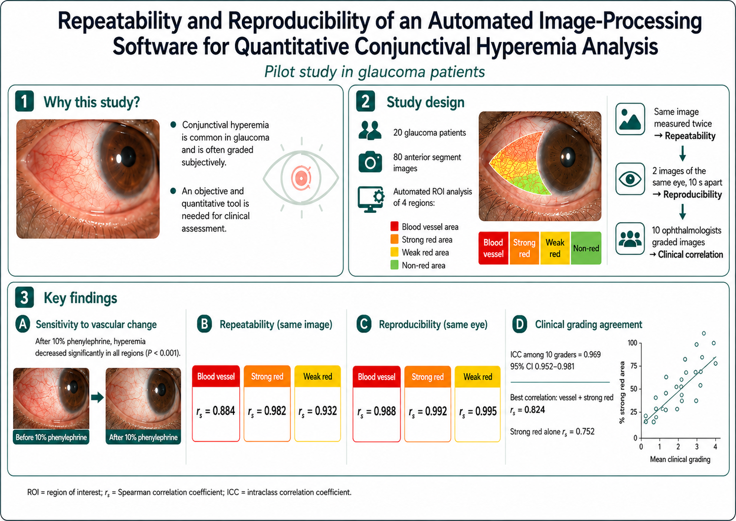
Objective Conjunctival hyperemia reflects the severity of various ocular conditions and is a common side effect of glaucoma medications, posing a significant concern for affected patients. Most existing clinical grading systems are subjective and rely heavily on individual physician interpretation, making interobserver comparison difficult. We developed an automated grading software to objectively and quantitatively assess conjunctival hyperemia. This study aimed to evaluate the repeatability and reproducibility of a newly developed automated software for quantitative assessment of conjunctival hyperemia in glaucoma patients in a pilot validation study.
Methods In this pilot study, glaucoma patients using at least one topical anti-glaucoma medication and exhibiting conjunctival hyperemia were enrolled. Each patient’s conjunctival hyperemia was assessed using the automated software, which segmented images into four regions: blood vessel area, strong red area, weak red area, and non-red area. An automatic region of interest (ROI) registration function was incorporated to enhance measurement consistency. Repeatability was assessed using two measurements from the same image, and reproducibility was tested using separate images of the same eye.
Results Eighty images from twenty glaucoma patients were analyzed (collected between December 1, 2016, and December 31, 2018). Following administration of 10% phenylephrine ophthalmic solution, the percentage
of conjunctival hyperemia decreased significantly in all regions (p < 0.001). Spearman correlation coefficients for repeated measurements on the same image were 0.884 (blood vessel), 0.982 (strong red), and 0.932 (weak red). Corresponding values for repeated images of the same eye were 0.988, 0.992, and 0.995, respectively. The intraclass correlation coefficient (ICC) for clinical grading by ten ophthalmologists was 0.969 (95% CI: 0.952–0.981).
Conclusion This pilot study demonstrates that the proposed automated software provides an objective and quantitative assessment of conjunctival hyperemia with excellent repeatability and reproducibility. The system shows high correlation with clinical grading and may serve as a reliable tool for monitoring conjunctival hyperemia changes in clinical practice.
References
Lindquist T. Conjunctivitis an overview and classification. In: Krachmer J, Mannis M, Holland E, editors. Cornea. 3rd ed. St. Louis: Mosby; 2010. p. 509–89.
Efron N. Grading scales for contact lens complications. Ophthalmic Physiol Opt. 1998;18:182–6.
Efron N, Pritchard N, Brandon K, Copeland J, Godfrey R, Hamlyn B, et al. A survey of the use of grading scales for contact lens complications in optometric practice. Clin Exp Optom. 2011;94:193–9.
Terry RL, Schnider CM, Holden BA, Cornish R, Grant T, Sweeney D, et al. CCLRU standards for success of daily and extended wear contact lenses. Optom Vis Sci. 1993;70:234–43.
Wolffsohn JS, Naroo SA, Christie C, Morris J, Conway R, Maldonado-Codina C; British Universities Committee of Contact Lens Educators (BUCCLE). Anterior eye health recording. Cont Lens Anterior Eye. 2015;38: 266–71.
Schulze MM, Jones DA, Simpson TL. The development of validated bulbar redness grading scales. Optom Vis Sci. 2007;84:976–83.
Fieguth P, Simpson T. Automated measurement of bulbar redness. Invest Ophthalmol Vis Sci. 2002;43: 340–7.
Yoneda T, Sumi T, Takahashi A, Hoshikawa Y, Kobayashi M, Fukushima A. Automated hyperemia analysis software: reliability and reproducibility in healthy subjects. Jpn J Ophthalmol. 2012;56:1–7.
Villumsen J, Ringquist J, Alm A. Image analysis of conjunctival hyperemia. A personal computer based system. Acta Ophthalmol (Copenh). 1991;69:536–9.
Efron N, Morgan PB, Jagpal R. Validation of computer morphs for grading contact lens complications. Ophthalmic Physiol Opt. 2002;22:341–9.
Dogan S, Astvatsatourov A, Deserno TM, Bock F, Shah-Hosseini K, Michels A, et al. Objectifying the conjunctival provocation test: photography-based rating and digital analysis. Int Arch Allergy Immunol. 2014;163:59–68.
Sárándi I, Claßen DP, Astvatsatourov A, Pfaar O, Klimek L, Mösges R, et al. Quantitative conjunctival provocation test for controlled clinical trials. Methods Inf Med. 2014;53:238–44.
Pfaar O, Claßen DP, Astvatsatourov A, Klimek L, Mösges R. Reliability of a new symptom score in a titrated quantitative conjunctival provocation test supported by an objective photodocumentation. Int Arch Allergy Immunol. 2018;176:215–24.
Pérez Bartolomé F, Martínez de la Casa JM, Arriola Villalobos P, Fernández Pérez C, Polo V, Sánchez Jean R, et al. Ocular redness measured with the Keratograph 5M in patients using anti-glaucoma eye drops. Semin Ophthalmol. 2018;33:643–50.
Wolffsohn JS. Incremental nature of anterior eye grading scales determined by objective image analysis. Br J Ophthalmol. 2004;88:1434–8.
Owen CG, Fitzke FW, Woodward EG. A new computer assisted objective method for quantifying vascular changes of the bulbar conjunctivae. Ophthalmic Physiol Opt. 1996;16:430–7.
Chen PC, Kovalcheck SW, Zweifach BW. Analysis of microvascular network in bulbar conjunctiva by image processing. Int J Microcirc Clin Exp. 1987;6:245–55.
Maldonado MJ, Arnau V, Martínez-Costa R, Navea A, Mico FM, Cisneros AL, et al. Reproducibility of digital image analysis for measuring corneal haze after myopic photorefractive keratectomy. Am J Ophthalmol. 1997;123:31–41.
Sumi T, Yoneda T, Fukuda K, Hoshikawa Y, Kobayashi M, Yanagi M, et al. Development of automated conjunctival hyperemia analysis software. Cornea. 2013;32_(Suppl_1):S52–9.
Huntjens B, Basi M, Nagra M. Evaluating a new objective grading software for conjunctival hyperaemia. Cont Lens Anterior Eye. 2020;43:137–43.
Schulze MM, Ng A, Yang M, Panjwani F, Srinivasan S, Jones LW, et al. Bulbar redness and dry eye disease: comparison of a validated subjective grading scale and an objective automated method. Optom Vis Sci. 2021;98:113–20.
Ostheimer P, Lins A, Helle LA, Romano V, Steger B, Augustin M, et al. Conjunctival Bulbar Redness Extraction Pipeline for High-Resolution Ocular Surface Photography. Transl Vis Sci Technol. 2025;14:6. PubMed PMID: 39786738
Li M, Huang K, Ma X, Wang Y, Wen F, Chen Q. Mask distillation network for conjunctival hyperemia severity classification. Mach Intell Res. 2023;20:909-22.
Hassanpour H, Samadiani N, Salehi SM. Using morphological transforms to enhance the contrast of medical images. Egypt J Radiol Nucl Med. 2015;46:481–9.
Huang SM. A study of hue identification in the hue circle of the HSB color space. Percept Mot Skills. 2005;100:1143-54.
Tuytelaars T, Mikolajczyk K. Local invariant feature detectors: a survey. Found Trends Comput Graph Vis. 2008;3:177–280.
Tareen SAK, Saleem Z. A comparative analysis of SIFT, SURF, KAZE, AKAZE, ORB, and BRISK. In: Computing, Mathematics and Engineering Technologies (iCoMET), 2018 IEEE International Conference on; 2018. p. 1–10.
Dalal N, Triggs B. Histograms of oriented gradients for human detection. In: CVPR 2005. IEEE Computer Society Conference on Computer Vision and Pattern Recognition; 2005. p. 886–93.
Vincent E, Laganière R. Detecting planar homographies in an image pair. In: Image and Signal Processing and Analysis, 2001 IEEE 2nd International Symposium on; 2001. p. 182–7.
Peterson RC, Wolffsohn JS. Sensitivity and reliability of objective image analysis compared to subjective grading of bulbar hyperaemia. Br J Ophthalmol. 2007; 91:1464–6.