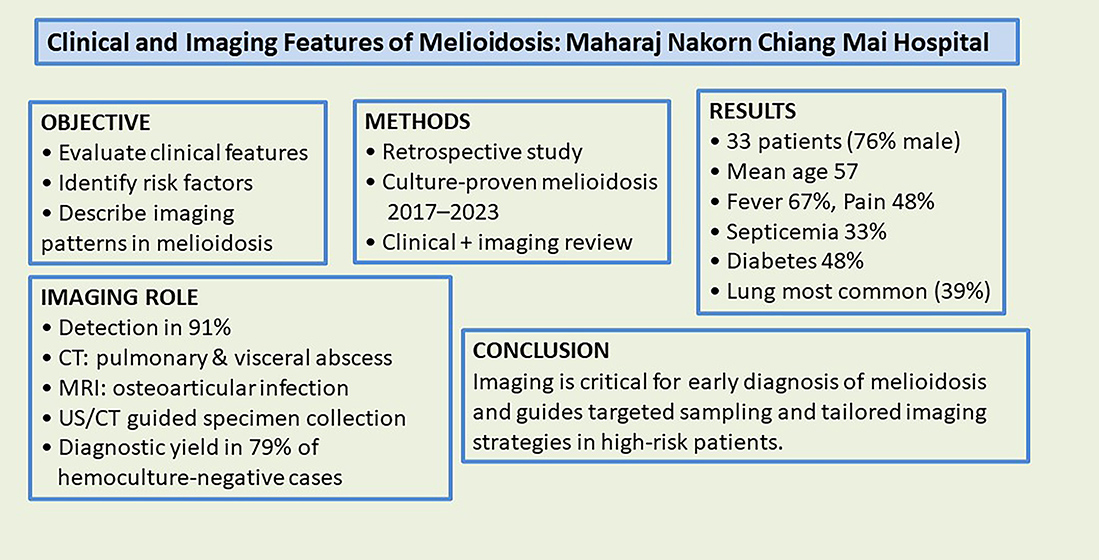
Clinical and Imaging Features of Melioidosis: A Study at Maharaj Nakorn Chiang Mai Hospital
DOI:
https://doi.org/10.12982/BSCM.2026.34Keywords:
melioidosis, sepsis, pneumonia, diagnostic imaging, multi- detector computed tomographyAbstract
Objective Melioidosis is a life-threatening infectious disease caused by Burkholderia pseudomallei. The clinical presentations are diverse with high mortality. The disease requires timely and appropriate antibiotics. Imaging studies play an important role in the diagnosis. This study evaluated clinical manifestations, risk factors, and imaging patterns of melioidosis.
Methods This retrospective study reviewed medical records and imaging studies of patients with melioidosis conclusively identified by cultures at Maharaj Nakorn Chiang Mai Hospital during 2017–2023.
Results Of 33 melioidosis patients, most were male (75.76%) and the mean age was 57. Fever (66.67%) and localized pain (48.48%) were the common symptoms, while septicemia occurred in 33.33% of cases. Diabetes mellitus (48.48%) was the predominant risk factor. Imaging studies, including chest radiographs, computed tomography (CT), magnetic resonance imaging (MRI), and ultrasonography, detected infections in 90.90% of the patients, with lung involvement being the most frequent (39.39%). CT was particularly effective in identifying pulmonary and visceral abscesses,
while MRI aided in diagnosing osteoarticular infections. Imaging successfully guided specimen collection in 78.57% of hemoculture-negative cases.
Conclusions This study underscores the importance of imaging in diagnosing melioidosis, particularly in patients with risk factors, and highlights the need for tailored imaging strategies based on clinical features.
References
Meumann EM, Limmathurotsakul D, Dunachie SJ, Wiersinga WJ, Currie BJ. Burkholderia pseudomallei and melioidosis. Nat Rev Microbiol. 2024;22:155-69.
Birnie E, Biemond JJ, Wiersinga WJ. Drivers of melioidosis endemicity: epidemiological transition, zoonosis, and climate change. Curr Opin Infect Dis. 2022;35:196-204.
Birnie E, Virk HS, Savelkoel J, Spijker R, Bertherat E, Dance DAB, et al. Global burden of melioidosis in 2015: a systematic review and data synthesis. Lancet Infect Dis. 2019;19:892-902.
Centers for Disease Control and Prevention. Melioidosis [Internet]. Atlanta: Centers for Disease Control and Prevention; [cited 2023 Aug 30]. Available from: https://ndc.services.cdc.gov/case-definitions/melioidosis-burkholderia-pseudomallei/.
Currie BJ. Melioidosis and Burkholderia pseudomallei : progress in epidemiology, diagnosis, treatment and vaccination. Curr Opin Infect Dis. 2022;35:517-23.
Gassiep I, Armstrong M, Norton R. Human Melioidosis. Clin Microbiol Rev. 2020;33:e00006-19.
Chakravorty A, Heath CH. Melioidosis: An updated review. Aust J Gen Pract. 2019;48:327-32.
Fairley L, Smith S, Maisrikrod S, Henning L. Systematic review and meta-analysis of diagnostic tests for diagnosis of melioidosis. Acta Trop. 2021;214:105784.
Raja NS, Scarsbrook C. Burkholderia Pseudomallei causing bone and joint infections: a clinical update. Infect Dis Ther. 2016;5:17-29.
Alsaif HS, Venkatesh SK. Melioidosis: spectrum of radiological manifestations. Saudi J Med Med Sci. 2016; 4:74-8.
Chong VF, Fan YF. The radiology of melioidosis. Australasian radiology. 1996;40:244-9.
Lim KS, Chong VH. Radiological manifestations of melioidosis. Clin Radiol. 2010;65:66-72.
Muttarak M, Peh WC, Euathrongchit J, Lin SE, Tan AG, Lerttumnongtum P, et al. Spectrum of imaging findings in melioidosis. The British journal of radiology. 2009;82:514-21.
Singcharoen T. CT findings in melioidosis. Australasian radiology. 1989;33:376-8.
Tan AP, Pui MH, Tan LK. Imaging patterns in melioidosis. Australasian Radiology. 1995;39:260-4.
Carrillo-Bayona JA, Alvarado-Benavides AM, Rodriguez JY, Alvarez-Moreno CA. Imaging manifestations of pulmonary melioidosis: A case series. Radiologia (Engl Ed). 2022;64:484-8.
Dhiensiri T, Puapairoj S, Susaengrat W. Pulmonary melioidosis: clinical-radiologic correlation in 183 cases in northeastern Thailand. Radiology. 1988;166:711-5.
Norman FF, Chen LH. Travel-associated melioidosis: a narrative review. J Travel Med. 2023;30:926.
Wu H, Wang X, Zhou X, Chen S, Mai W, Huang H, et al. Osteomyelitis and septic arthritis due to Burkholderia pseudomallei: A 10-year retrospective melioidosis study from south China. Front Cell Infect Microbiol. 2021;11:654745.
Currie BJ, Ward L, Cheng AC. The epidemiology and clinical spectrum of melioidosis: 540 cases from the 20 year Darwin prospective study. PLoS Negl Trop Dis. 2010;4:e900.
Yu A, Su L, Li Q, Li X, Tao S, Li F, et al. Imaging and clinical manifestations of hematogenous dissemination in melioidosis. BMC Med Imaging. 2024;24:296.
Khiangte HL, Vimala LR, Eapen A, Veeraraghavan B, Karuppusami R, Gibikote S. A retrospective case-control study to evaluate the diagnostic accuracy of honeycomb sign in melioid liver abscess. Am J Trop Med Hyg. 2018;99:852-7.
Pattamapaspong N, Muttarak M. Musculoskeletal melioidosis. Semin Musculoskelet Radiol. 2011;15:480-8.
Kozlowska J, Smith S, Roberts J, Pridgeon S, Hanson J. Prostatic abscess due to Burkholderia pseudomallei: facilitating diagnosis to optimize management. Am J Trop Med Hyg. 2018;98:227-30.
Morse LP, Moller CC, Harvey E, Ward L, Cheng AC, Carson PJ, et al. Prostatic abscess due to Burkholderia pseudomallei: 81 cases from a 19-year prospective melioidosis study. J Urol. 2009;182:542-7; discussion 7.
Wen SC, Juan YS, Wang CJ, Chang K, Shih MC, Shen JT, et al. Emphysematous prostatic abscess: case series study and review. Int J Infect Dis. 2012;16:e344-9.
Hirunpat P, Panyaping T, Wongpipathpong W, Hirunpat S. Imaging clues for the diagnosis of various pathogenic causes of infectious spondylitis. Skeletal Radiol.
;54:2411-24.
Lee SC, Ling TS, Chen JC, Huang BY, Sheih WB. Melioidosis with adrenal gland abscess. Am J Trop Med Hyg. 1999;61:34-6.
Teerajetgul S, Jantarapootirat M, Traiwanatham S, Kirdlarp S, Sriphrapradang C. Isolated adrenal abscess due to melioidosis. Am J Trop Med Hyg. 2025; 113:395-8.
Guo YK, Yang ZG, Li Y, Ma ES, Deng YP, Min PQ, et al. Addison’s disease due to adrenal tuberculosis: contrast-enhanced CT features and clinical duration correlation. Eur J Radiol. 2007;62:126-31.
Udare A, Agarwal M, Siegelman E, Schieda N. CT and MR imaging of acute adrenal disorders. Abdom Radiol (NY). 2021;46:290-302.
Meumann EM, Currie BJ. Approach to melioidosis. CMI Communications. 2024;1:100008. doi:10.1016/j.cmicom.2024.100008