Comparison of Contrast-Enhanced MRI Using Hepatocyte-Specific Contrast with Contrast-Enhanced CT in Evaluation of Hepatobiliary Space-Occupying Lesions
Keywords:
contrast-enhanced computed tomography, contrast-enhanced magnetic resonance imaging, diagnostic accuracy, lesion characterization, imaging modalitiesAbstract
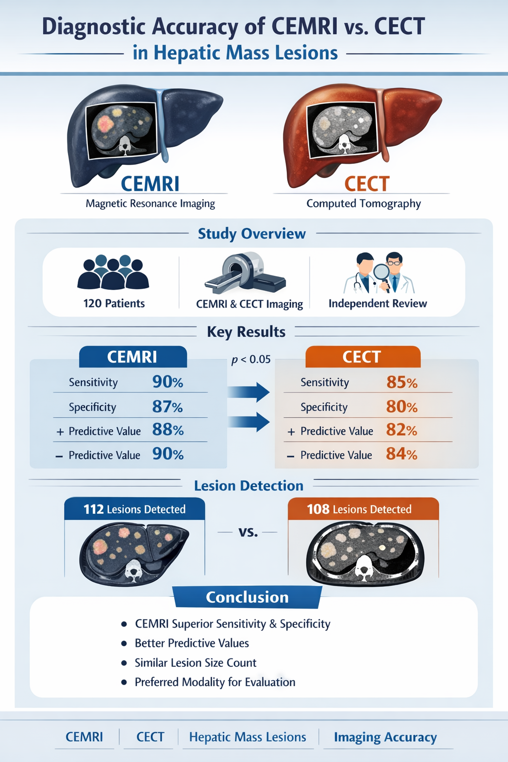
Objective This study aimed to evaluate the diagnostic accuracy of multiphasic contrast-enhanced computed tomography (CECT) and contrast- enhanced magnetic resonance imaging (CEMRI) in assessing hepatic mass lesions.
Methods A cohort of 120 patients underwent imaging with both CEMRI and CECT to compare their diagnostic capabilities. Findings were analyzed for lesion size, enhancement patterns, sensitivity, specificity, and predictive values. Statistical tests were conducted to assess the significance of differences between the modalities, with p-values reported for critical metrics. To ensure consistency and minimize bias, a standardized imaging protocol was followed, and radiology experts independently reviewed all images.
Results CEMRI demonstrated higher sensitivity (90%) and specificity (87%) compared to CECT (85% and 80%, respectively), with statistically significant differences (p < 0.05). CEMRI also outperformed CECT in posi-
tive predictive value (88% vs. 82%) and negative predictive value (90% vs. 84%). While the mean lesion size detected by CEMRI (4.0 ± 2.0 cm) was slightly smaller than that detected by CECT (4.4 ± 2.2 cm), the difference was not statistically significant (p = 0.45). Additionally, CEMRI identified 112 lesions compared to 108 lesions detected by CECT, further highlighting its superior diagnostic accuracy, particularly in detecting heterogeneous lesions.
Conclusion CEMRI outperforms CECT in diagnostic sensitivity, specificity, and predictive values, making it the preferred modality for lesion detection and characterization. However, both modalities performed similarly in assessing lesion size and total lesion count. These findings underscore the value of CEMRI in clinical scenarios requiring detailed lesion evaluation. Future studies with larger cohorts and a broader range of pathologies are recommended to confirm and extend these results for greater clinical applicability.
References
Klempnauer J, Ridder GJ, Werner M, Weimann A, Pichlmayr R. What constitutes long-term survival after surgery for hilar cholangiocarcinoma? Cancer. 1997;79:26–34.
Nagakawa Y, Kasuya K, Bunso K, Hosokawa Y, Kuwabara H, Nakagima T, et al. Usefulness of multi-
-dimensional computed tomograms fused with multiplanar reconstruction images and peroral cholangioscopy findings in hilar cholangiocarcinoma. J Hepato-Biliary-Pancreat Sci. 2014;21:256–62.
Viera AJ, Garrett JM. Understanding interobserver agreement: the kappa statistic. Fam Med. 2005;37:360-3.
Sakamoto E, Nimura Y, Hayakawa N, Kamiya J, Kondo S, Nagino M, et al. The pattern of infiltration at the proximal border of hilar bile duct carcinoma: a histologic analysis of 62 resected cases. Ann Surg. 1998; 227:405–11.
Ruys AT, van Beem BE, Engelbrecht MRW, Bipat S, Stoker J, Van Gulik TM. Radiological staging in patients with hilar cholangiocarcinoma: a systematic review and meta-analysis. Br J Radiol. 2012;85:1255–62.
Lee DH, Kim B, Lee ES, Kim HJ, Min JH, Lee JM, et al. Radiologic evaluation and structured reporting form for extrahepatic bile duct cancer: 2019 consensus recommendations from the Korean Society of Abdominal Radiology. Korean J Radiol. 2021;22:41–62.
Park HS, Lee JM, Choi JY, Lee MW, Kim HJ, Han JK, et al. Preoperative evaluation of bile duct cancer: MRI combined with MR cholangiopancreatography versus MDCT with direct cholangiography. AJR Am J Roentgenol. 2008;190:396–405.
Lee HY, Kim SH, Lee JM, Kim SW, Jang JY, Han JK, et al. Preoperative assessment of resectability of hepatic hilar cholangiocarcinoma: combined CT and cholangiography with revised criteria. Radiology. 2006;239: 113–21.
Ryoo I, Lee JM, Chung YE, Park HS, Kim SH, Han JK, et al. Gadobutrol-enhanced, three-dimensional, dynamic MR imaging with MR cholangiography for the preoperative evaluation of bile duct cancer. Invest Radiol. 2010;45:217–24.
Lee DH, Kim B, Lee JM, Lee ES, Choi MH, Kim H. Multidetector CT of extrahepatic bile duct cancer: diagnostic performance of tumor resectability and interreader agreement. Radiology. 2022;304:96–105.
Franken LC, Coelen RJS, Erdmann JI, Verheij J, Kop MP, van Gulik TM, et al. Multidetector computed tomography assessment of vascular involvement in perihilar cholangiocarcinoma. Quant Imaging Med Surg. 2021; 11:4514–21.
Rassam F, Roos E, van Lienden KP, van Hooft JE, Klümpen HJ, van Tienhoven G, et al. Modern work-up and extended resection in perihilar cholangiocarcinoma: the AMC experience. Langenbecks Arch Surg. 2018;403:289–307.
Raghavan K, Jeffrey RB, Patel BN, DiMaio MA, Willmann JK, Olcott EW. MDCT diagnosis of perineural invasion involving the celiac plexus in intrahepatic cholangiocarcinoma: preliminary observations and clinical implications. AJR Am J Roentgenol. 2015;205: W578-584.
Ni Q, Wang H, Zhang Y, Qian L, Chi J, Liang X, et al. MDCT assessment of resectability in hilar cholangiocarcinoma. Abdom Radiol N Y. 2017;42:851–60.
Takayasu K, Moriyama N, Muramatsu Y, Shima Y, Goto H, Yamada T. Intrahepatic portal vein branches studied by percutaneous transhepatic portography. Radiology. 1985;154:31–6.
Wang R, Liu Q, Zhang W. Coping, social support, and family quality of life for caregivers of individuals with autism: Meta-analytic structural equation modeling. Personal Individ Differ. 2022;186:111351.
Sica GT, Ji H, Ros PR. Computed tomography and magnetic resonance imaging of hepatic metastases. Clin Liver Dis. 2002;6:165–79.
Coenegrachts K. Magnetic resonance imaging of the liver: New imaging strategies for evaluating focal liver lesions. World J Radiol. 2009;1:72.
Choi JY, Lee JM, Sirlin CB. CT and MR imaging diagnosis and staging of hepatocellular carcinoma: part II. extracellular agents, hepatobiliary agents, and ancillary imaging features. Radiology. 2014;273:30–50.
Kim YY, Lee S, Shin J, Son WJ, Roh YH, Hwang JA, et al. Diagnostic performance of CT versus MRI liver imaging reporting and data system category 5 for hepatocellular carcinoma: a systematic review and meta-analysis of comparative studies. Eur Radiol. 2022; 32:6723–9.
Vogl TJ, Kümmel S, Hammerstingl R, Schellenbeck M, Schumacher G, Balzer T, et al. Liver tumors: comparison of MR imaging with Gd-EOB-DTPA and Gd-DTPA. Radiology. 1996;200:59–67.
Thian YL, Riddell AM, Koh DM. Liver-specific agents for contrast-enhanced MRI: role in oncological imaging. Cancer Imaging Off Publ Int Cancer Imaging Soc. 2013;13:567–79.