
Deep learning approaches for CT image reconstruction: Advancements and clinical implications
Article Sidebar
Main Article Content
Abstract
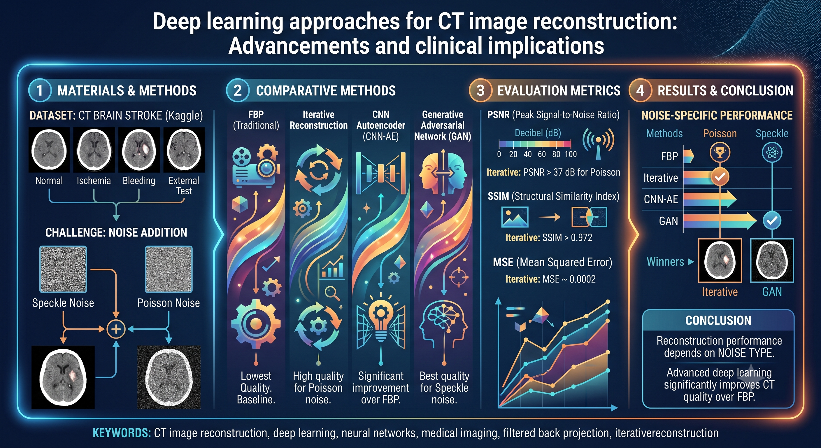
Background: CT is a trusted imaging modality for diagnosing disease. The quality of the CT image plays the most important role in disease prediction. Images will be degraded by noise, blur, and other artifacts. Therefore, the enhancement and reconstruction of CT images are required before processing for prediction. Enhancements are only cosmetic changes in images, but reconstruction provides well-defined models for reconstruction. In this paper, we have compared the performance of these four CT brain reconstruction and enhancement techniques: Filtered back projection (FBP), Iterative reconstruction, Generative Adversarial Network (GAN), and CNN autoencoder, by adding Speckle and Poisson noise.
Objectives: The possible objective statements of this study will cover a broad spectrum of developments in medical imaging technology. These are aimed at enhancing diagnostic test quality, patient safety, and overall care outcomes through innovative imaging methods and data analysis. The suggested objectives involve integrating an artificial intelligence algorithm to enhance them, thereby improving patient care and diagnostic accuracy across numerous medical fields. In this experiment, the CT brain stroke dataset was obtained from the Kaggle database and evaluated using performance metrics (PSNR, SSIM, and MSE) across the normal, ischemia, bleeding, and external test folders, with Poisson and Speckle noise added using the GAN, CNN autoencoder, iterative, and FBP techniques.
Materials and methods: The CT brain stroke dataset contains CT images organized in four folders: normal, ischemia, bleeding, and external test. These folders can be used to evaluate performance metrics. The methods applied include: Convolutional Neural Network (CNN) autoencoder, Generative Adversarial Network (GAN), iterative reconstruction, and FBP, each implemented with hyper-tuned parameters. Experimental results calculate PSNR, SSIM, and MSE after adding Poisson and Speckle noise to test the above deep learning algorithms.
Results: The lowest image quality was achieved for FBP for all datasets and both noise types. When the noise was Poisson, the performance of iterative reconstruction was the best, with PSNR exceeding 37 dB, SSIM exceeding 0.972, and MSE as low as 0.0002. On adding Poisson noise, the GAN achieved higher PSNR values and SSIM values up to 32.54 dB and 0.938, respectively, and with low errors in reconstruction, except for the external test folder. The CNN autoencoder (CNN-AE) also yielded significant improvements over FBP, achieving competitive PSNR and SSIM scores across all datasets.
Conclusion: The results indicate that the quality of CT images can be significantly improved using advanced deep learning and Iterative reconstruction methods compared with conventional FBP. It is observed that iterative reconstruction yields the best results for images corrupted by Poisson noise, whereas GANs perform best for images corrupted by Speckle noise. Therefore, reconstruction performance depends on the nature of the noise present in the CT images.
Article Details

This work is licensed under a Creative Commons Attribution-NonCommercial-NoDerivatives 4.0 International License.
Personal views expressed by the contributors in their articles are not necessarily those of the Journal of Associated Medical Sciences, Faculty of Associated Medical Sciences, Chiang Mai University.
References
Zhang M, Shi Y, & Gu S. The use of deep learning methods in low-dose computed tomography image reconstruction: a systematic review. Complex Intell Syst. 2022; 8(6): 5545-61. doi: 10.1007/s40747-022-00724-7.
Fortuni F, Franchin L, Gabrielli D, Vitale E, Conte E, Spinelli A, et al. Advancements and applications of artificial intelligence in cardiovascular imaging: a comprehensive review. Eur Heart J Imaging Methods Pract. 2024; 2(4). doi: 10.1093/ehjimp/qyae136.
Li Z, Tang C, Yan B, Cai A, Zhang W, Liang N, et al. Promising generative adversarial network based sinogram inpainting method for ultra-limitedangle computed tomography imaging. Sensors. 2019; 19(18): 3941. doi: 10.3390/s19183941.
Immonen E, Wong J, Nieminen M, Kekkonen L, Roine S, Törnroos S, et al. The use of deep learning towards dose optimization in low-dose computed tomography: a scoping review. Radiography (Lond). 2021; 28(1): 208-14. doi: 10.1016/j.radi.2021.07.010.
Mese I, Demirsoy U, AltintasMese C, & Anik Y. Innovative advances in pediatric radiology: computed tomography reconstruction techniques, photon-counting detector computed tomography, and beyond. Pediatr Radiol. 2023; 54(1): 1-11. doi: 10.1007/s00247-023-05823-2.
Mitra U, & Rehman SU. Leveraging AI and machine learning for next-generation clinical decision support systems (CDSS; pp 83-112). IGI Global. doi: 10.4018/979-8-3693-7277-7.ch003.
Southard RN, Martinot CA, Temkit MH, Augustyn RA, Bardo DME, & Thorkelson MA. Comparison of iterative model reconstruction versus filtered back-projection in pediatric emergency head CT: dose, image quality, and image-reconstruction times. AJNR Am J Neuroradiol. 2019; 40(5): 866-71. doi: 10.3174/ajnr.a6034.
Pontana F, Faivre JB, Remy-Jardin M, Hachulla AL, Duhamel A, Pagniez J, et al. Chest computed tomography using iterative reconstruction vs filtered back projection (Part 2): image quality of low-dose CT examinations in 80 patients. Eur Radiol. 2010; 21(3): 636-43. doi: 10.1007/s00330-010-1991-4.
Shi L, Karellas A, Vedantham S, & Zhu L. Library based x-ray scatter correction for dedicated cone beam breast CT. Med Phys. 2016; 43(8): 4529-44. doi: 10.1118/1.4955121.
Terzis R, Reimer RP, Nelles C, Celik E, Caldeira L, Heidenreich A, et al. Deep-learning-based image denoising in imaging of urolithiasis: assessment of image quality and comparison to state-of-theart iterative reconstructions. Diagnostics. 2023; 13(17): 2821. doi: 10.3390/diagnostics13172821.
Sadia RT, Zhang J, & Chen J. CT image denoising methods for image quality improvement and radiation dose reduction. J Appl Clin Med Phys. 2024; 25(2). doi: 10.1002/acm2.14270.
Radiuk P. Applying 3D U-Net architecture to the task of multi-organ segmentation in computed tomography. Appl Comput Syst. 2020; 25(1): 43-50. doi: 10.2478/acss-2020-0005.
Han C, Furukawa Y, Nakayama H, Hayashi H, Araki R, Rundo L, et al. Infinite brain MR images: PGGANbased data augmentation for tumor detection (Vol. 151; pp 291-303). Springer Singapore. doi: 10.1007/978-981-13-8950-4_27.
Lavudiya NS, & Prasad CVPR. Enhancing ophthalmological diagnoses: an adaptive ensemble learning approach using fundus and OCT imaging. Int J Comput Exp Sci Eng. 2024; 10(4). doi: 10.22399/ijcesen.678.
Hasan AM, Kahtan H, Al-Ahmad AS, Jalab HA, & Meziane F. Combining deep and handcrafted image features for MRI brain scan classification. IEEE Access. 2019; 7: 79959-67. doi: 10.1109/access.2019.292269.
Rahman H, Khan AR, Sadiq T, Farooqi AH, Khan IU, & Lim WH. A systematic literature review of 3D deep learning techniques in computed tomography reconstruction. Tomography. 2023; 9(6): 2158-89. doi: 10.3390/tomography9060169.
Liu X, Gu W, Yao T, Su S, Mo Y, & Shen J. Superresolution reconstruction of CT images based on multi-scale information fused generative adversarial networks. Ann Biomed Eng. 2023; 52(1): 57-70. doi: 10.1007/s10439-023-03412-w.
Ohashi K, Nagatani Y, Yoshigoe M, Iwai K, Tsuchiya K, Hino A, et al. Applicability evaluation of fullreference image quality assessment methods for computed tomography images. J Digit Imaging. 2023; 36(6): 2623-34. doi: 10.1007/s10278-023-00875-0.
Bornet PA, Blum A, Germain E, Gondim Teixeira PA, Lombard C, Villani N, et al. Clinical acceptance of deep learning reconstruction for abdominal CT imaging: objective and subjective image quality and low-contrast detectability assessment. Eur Radiol. 2022; 32(5): 3161-72. doi: 10.1007/s00330-021-08410-x.
Parakh A, Cao J, Kambadakone AR, Blake MA, Pierce TT, & Savage CA. Sinogram-based deep learning image reconstruction technique in abdominal CT: image quality considerations.Eur Radiol. 2021; 31(11): 8342-53. doi: 10.1007/s00330-021-07952-4.
Koç U, Akçapınar Sezer E, Alper Özkaya Y, et al. Artificial intelligence in healthcare competition (TEKNOFEST-2021): stroke data set. Eurasian J Med. 2022; 54(3): 248-58.
Nageswara Rao B, Acharya UR, Tan RS, Dash P, Mohapatra M, Sabut S. Convolutional autoencoder-based deep learning for intracerebral hemorrhage classification using brain CT images. Cogn Neurodyn. 2025 Dec; 19(1): 77.
Laino ME, Cancian P, Politi LS, Della Porta MG, Saba L, Savevski V. Generative adversarial networks in brain imaging: a narrative review. J Imaging. 2022 Mar 23; 8(4): 83.
Li Q, Li S, Li R, Wu W, Dong Y, Zhao J, et al. Lowdose computed tomography image reconstruction via a multistage convolutional neural network with autoencoder perceptual loss network. Quant Imaging Med Surg. 2022 Mar; 12(3): 1929.
Runkel C, Biguri A, Schönlieb CB. Continuous learned primal dual. In: 2024 IEEE 34th International Workshop on Machine Learning for Signal Processing (MLSP). 2024 Sep 22; pp 1-6. IEEE.
