
Method comparison and clinical classification agreement between Freelite and N-Latex serum-free light chain assays for clinical laboratory implementation
Article Sidebar
Main Article Content
Abstract
Background: Measurement of serum free light chains (SFLC) is an essential component in the diagnosis and monitoring of plasma cell dyscrasias. Two analytical platforms are widely used in clinical laboratories: the Freelite assay, which employs polyclonal antibodies with turbidimetric detection, and the N-Latex assay, which uses monoclonal antibodies with nephelometric measurement. Because these assays differ in antibody design, calibration, and analytical detection principles, numerical concentrations and κ/λ ratios may
not be directly interchangeable across platforms. This study aimed to evaluate the analytical agreement and clinical classification concordance between the Freelite and N-Latex assays to assess the feasibility of implementing the N-Latex method in routine laboratory practice.
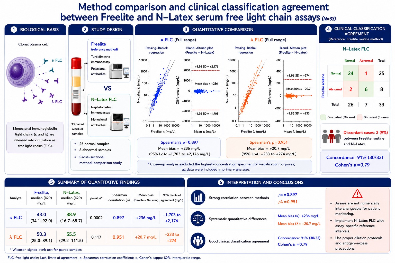
Materials and methods: A cross-sectional method-comparison study was performed using 33 paired archived residual serum samples obtained from routine clinical testing. Free κ and λ concentrations were measured using both Freelite and N-Latex assays on the same specimens. Analytical agreement was assessed using the Wilcoxon signed-rank test, Spearman correlation, Passing–Bablok regression, and Bland-Altman analysis. κ/λ ratios were interpreted according to assay-specific reference intervals, and categorical agreement was evaluated using Cohen’s kappa coefficient.
Results: Freelite produced significantly higher κ concentrations than N-Latex (median 43.0 vs 38.9 mg/L, p=0.0002), whereas λ concentrations did not differ significantly. Strong correlations were observed for both κ (ρ=0.897) and λ (ρ=0.951); however, correlation alone was not sufficient to demonstrate quantitative interchangeability. Passing-Bablok regression demonstrated proportional bias for κ measurements, with Freelite yielding higher values at elevated concentrations and the 95% confidence interval of the slope not including 1.0. Bland-Altman analysis revealed wide limits of agreement for κ values at extreme concentrations, indicating clinically relevant numerical differences between methods. When κ/λ ratios were interpreted categorically, using assay-specific reference intervals, concordant classification was observed in 30 of 33 samples (91%), corresponding to substantial agreement (κ=0.79).
Conclusions: The Freelite and N-Latex assays demonstrate strong correlation but exhibit systematic quantitative differences, particularly for κ measurements at high concentrations. Although κ/λ ratio interpretation is largely concordant, the two assays should not be considered directly interchangeable for absolute numerical interpretation or longitudinal monitoring. Implementation of the N-Latex assay is feasible in routine clinical laboratories provided that assay-specific reference intervals, appropriate dilution protocols, and antigen-excess precautions are applied, and clinicians are informed about expected differences when transitioning between
analytical platforms.
Article Details

This work is licensed under a Creative Commons Attribution-NonCommercial-NoDerivatives 4.0 International License.
Personal views expressed by the contributors in their articles are not necessarily those of the Journal of Associated Medical Sciences, Faculty of Associated Medical Sciences, Chiang Mai University.
References
Dispenzieri A, Kyle RA, Katzmann JA, Therneau TM, Larson DR, Benson JT, et al. Immunoglobulin free light chain ratio is an independent risk factor for progression of monoclonal gammopathy of undetermined significance. Leukemia. 2009; 23(2): 215-3. doi: 10.1038/leu.2008.311.
Rajkumar SV, Dimopoulos MA, Palumbo A, Blade J, Merlini G, Mateos MV, et al. International Myeloma Working Group updated criteria for the diagnosis of multiple myeloma. Lancet Oncol. 2014; 15(12): e538-48. doi: 10.1016/S1470-2045(14)70442-5.
Bradwell AR, Carr-Smith HD, Mead GP, Tang LX, Showell PJ, Drayson MT, et al. Highly sensitive, automated immunoassay for immunoglobulin free light chains in serum and urine. Clin Chem Lab Med. 2003; 41(3): 321-30. doi: 10.1515/CCLM.2003.053.
Kastritis E, Terpos E, Roussou M, Gavriatopoulou M, Migkou M, Pamboucas C, et al. Evaluation of the prognostic value of serum free light chain ratio in patients with symptomatic multiple myeloma. Leukemia. 2015; 29(6): 1337-44. doi: 10.1038/leu.2015.42.
Wu AH. Performance of immunoassays for detection of free light chains in serum and urine. Clin Chim Acta. 2014; 431: 1-7. doi: 10.1016/j.cca.2014.01.025.
Tosi P, Tomassetti S, Merli A, Polli V. Serum free light-chain assay for the detection and monitoring of multiple myeloma and related conditions. Ther Adv Hematol. 2013; 4(1): 37-41. doi: 10.1177/2040620712466863.
Tate JR, Mollee P, Dimeski G, Carter AC, Gill D. Analytical performance of serum free light-chain assay during monitoring of multiple myeloma. Clin Chem. 2012; 58(3): 523-39. doi: 10.1373/clinchem.2011.170662.
Te Velthuis H, Knop I, Stam P, van den Bergh FA, Ooms B, Smit JW, et al. N Latex FLC - new monoclonal high-performance assays for the determination of free light chain kappa and lambda. Clin Chem. 2011; 57(4): 499-506. doi: 10.1373/clinchem.2010.155820.
Palladini G, Merlini G. Serum free light chain assay for monitoring patients with AL amyloidosis. Blood. 2014; 124(4): 535-43. doi: 10.1182/blood-2014-03-535385.
Ladwig PM, Barnidge DR, Snyder MR, Katzmann JA, Murray DL. Quantification of serum kappa free light chains using two different assays: Assessment of precision, limit of quantification, and biological variation. Am J Clin Pathol. 2014; 142(2): 231-9. doi: 10.1309/AJCPOC7U9RCHYVLI.
Zegers I, Van der Heyden S, Sciacovelli L, Secchiero S, Brandslund I, Streichert T, et al. Towards a reference system for serum free light chain measurements. Clin Chem Lab Med. 2019; 57(9): 1301-7. doi: 10.1515/cclm-2019-0145.
Svarstad E, Bostad L, Myklebust R, Harboe T, Hovig T, Langholm R, et al. Light chain nephropathy in patients with multiple myeloma: A clinicopatho logical study of 41 patients. Nephrol Dial Transplant. 2013; 28(5): 1234-41. doi: 10.1093/ndt/gfs470.
Jacobs JF, Hoedemaekers R, Teunesen MA, Willems HL, van den Bergh FA, van der Hoeven JG. Serum free light chain analysis: Reference values in healthy children. Clin Chem. 2011; 57(5): 713-20. doi: 10.1373/clinchem.2010.160135.
Miller WG, Tate JR, Barth JH, Jones GR, Parslow RA, Rifai N, et al. Harmonization of laboratory testing-current achievements and future strategies. Arch Pathol Lab Med. 2014; 138(2): 234-43. doi: 10.5858/arpa.2013-0268-CP.
Clinical and Laboratory Standards Institute (CLSI). Measurement Procedure Comparison and Bias Estimation Using Patient Samples; Approved Guideline - Third Edition (EP09-A3). Wayne, PA: CLSI; 2013.
Hutchison CA, Harding S, Hewins P, Mead GP, Townsend J, Bradwell AR, et al. Quantitative assessment of serum and urinary polyclonal free light chains in patients with chronic kidney disease. Clin J Am Soc Nephrol. 2008; 3(6): 1684-90. doi: 10.2215/CJN.02290508.
Lippi G, Avanzini P, Pavesi F, Aloe R, Cervellin G. Evaluation of serum free light chains in patients with chronic kidney disease. Clin Chim Acta. 2017; 464: 19-25. doi: 10.1016/j.cca.2016.11.016.
Abraham RS, Clark RJ, Bryant S, Larson T, Kyle RA, Katzmann JA. Correlation of serum immuno globulin free light chain quantification with urinary Bence Jones protein in light chain myeloma. Arch Pathol Lab Med. 2012; 136(5): 563-72. doi: 10.5858/arpa.2011-0317-OA.
Ho C, Pacaud L, Evison F, Pratt G, Harding S, Zaihra T, et al. Multicenter evaluation of the N Latex FLC assay for the detection of monoclonal free light chains and comparison with Freelite and serum protein electrophoresis. Diagnostics (Basel). 2021; 11(8): 1416. doi: 10.3390/diagnostics11081416.
Daves M, Giacomuzzi F, Bergmann M, Cemin R. Analytical comparison of Freelite™ and N Latex FLC assays for quantifying serum free light chains and impact on the diagnosis of monoclonal gammopathies. Clin Chem Lab Med. 2021; 59(1): 117-24. doi: 10.1515/cclm-2020-0585.
Sasson SC, Lawrie C, Rivory LP, Ho PJ. Comparison of two serum free light chain assays for the diagnosis and monitoring of patients with plasma cell dyscrasias. Intern Med J. 2015; 45(1): 60-6. doi: 10.1111/imj.12624.
