
Assessment of imaging radiation dose from tomotherapy systems
Article Sidebar
Main Article Content
Abstract
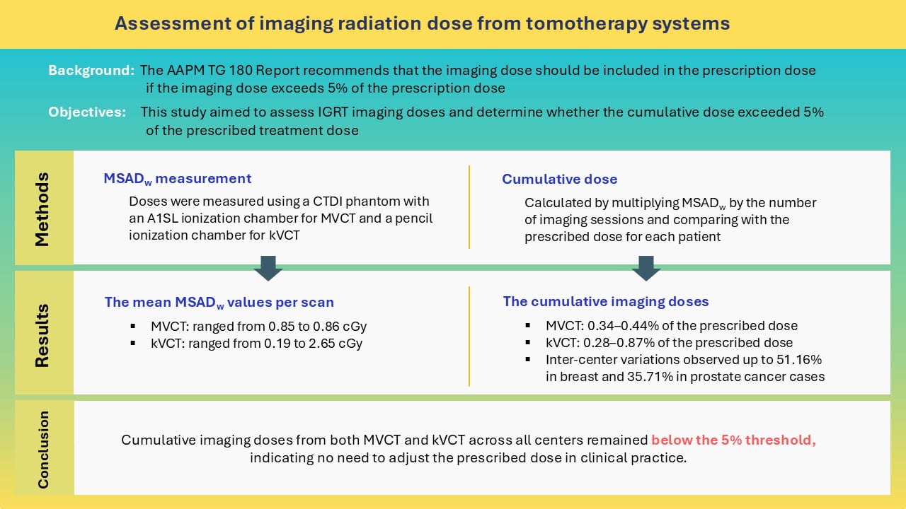
Background: Tomotherapy systems employ image-guided radiotherapy (IGRT) using either megavoltage (MVCT) or kilovoltage (kVCT) computed tomography for daily patient positioning verification. However, these imaging procedures can deliver a non-negligible radiation dose to the patient. According to the American Association of Physicists in Medicine (AAPM) Task Group 180, this dose should be incorporated into the prescribed treatment dose if it exceeds 5%.
Objectives: This study aimed to assess IGRT imaging doses and determine whether the cumulative dose exceeded 5% of the prescribed treatment dose.
Materials and methods: Imaging doses from IGRT were evaluated under clinical scan protocols across four tomotherapy machines at three centers, including two MVCT systems and two kVCT systems. Doses were measured as Weighted Multiple Slice Average Dose (MSADw) using a CTDI phantom. Cumulative imaging dose was calculated by multiplying MSADw by the number of imaging sessions, then compared to the prescribed dose for each patient.
Results: The mean MSADw values for MVCT ranged from 0.85 to 0.86 cGy per scan, while for kVCT it ranged from 0.19 to 2.65 cGy per scan. Over the treatment course, cumulative imaging doses accounted for 0.34-0.44% of the prescribed dose for MVCT and 0.28-0.87% for kVCT. Notable inter-center variations were observed, with differences of up to 51.16% in breast cancer cases and 35.71% in prostate cancer cases.
Conclusion: Cumulative imaging doses from both MVCT and kVCT across all centers remained below the 5% threshold, indicating no need to adjust the prescribed dose in clinical practice. Nevertheless, further studies are required to identify the causes of inter-center variations and to develop optimized imaging protocols aimed at minimizing patient dose while maintaining adequate image quality.
Article Details

This work is licensed under a Creative Commons Attribution-NonCommercial-NoDerivatives 4.0 International License.
Personal views expressed by the contributors in their articles are not necessarily those of the Journal of Associated Medical Sciences, Faculty of Associated Medical Sciences, Chiang Mai University.
References
Tegtmeier RC, Ferris WS, Bayouth JE, Miller JR, Culberson WS. Characterization of imaging performance of a novel helical kVCT for use in image-guided and adaptive radiotherapy. J Appl Clin Med Phys. 2022; 23: e13648. doi.org/10.1002/acm2.13648.
Yang B, Geng H, Chang TYA, Tse MY, Lam WW, Huang CY, et al. Clinical implementation of kVCT-guided tomotherapy with ClearRT. Phys Eng Sci Med. 2022; 45: 915-24. doi.org/10.1007/s13246-022-01162-y.
Sakai Y, Monzen H, Takei Y, Kosaka H, Nakamura K, Yanagi Y, et al. Evaluation of in-room volumetric imaging doses for image-guided radiotherapy: A multi-institutional study. J Med Phys. 2023; 48: 189-94. doi.org/10.4103/jmp.jmp_109_22.
Tomita T, Isobe T, Furuyama Y, Takei H, Kobayashi D, Mori Y, et al. Evaluation of dose distribution and normal tissue complication probability of a combined dose of cone-beam computed tomography imaging with treatment in prostate intensity-modulated radiation therapy. J Med Phys. 2020; 45: 78-87. doi.org/10.4103/jmp.JMP_4_20.
Brenner DJ. Induced second cancers after prostate-cancer radiotherapy: No cause for concern? Int J Radiat Oncol Biol Phys. 2006; 65: 637-9. doi.org/10.1016/j.ijrobp.2006.02.044.
Ding GX, Alaei P, Curran B, Flynn R, Gossman M, Mackie TR, et al. Image guidance doses delivered during radiotherapy: Quantification, management, and reduction: Report of the AAPM Therapy Physics Committee Task Group 180. Med Phys. 2018; 45:e84-9. doi.org/10.1002/mp.12824.
Dische S, Saunders MI, Williams C, Hopkins A, Aird E. Precision in reporting the dose given in a course of radiotherapy. Rad Onc.1993; 29: 287-93. doi.org/10.1016/0167-8140(93)90146-Y.
Saunders MI, Dische S, Grosch EJ, Fermont DC, Ashford RFU, Maher EJ, et al. Experience with CHART. Int J Radiat Oncol Biol Phys. 1991; 21: 871-8. doi. org/10.1016/0360-3016(91)90722-G.
Morgan-Fletcher SL. Prescribing, Recording and Reporting Photon Beam Therapy (Supplement to ICRU Report 50), ICRU Report 62. ICRU, pp. ix+52, 1999 (ICRU Bethesda, MD) $65.00 ISBN 0-913394-61-0. Br J Radiol. 2001; 74: 294. doi.org/10.1259/bjr.74.879.740294.
Langen KM, Papanikolaou N, Balog J, Crilly R, Followill D, Goddu SM, et al. QA for helical tomotherapy: Report of the AAPM Task Group 148. Med Phys. 2010; 37: 4817-53. doi.org/10.1118/1.3462971.
Mege JP, Wenzhao S, Veres A, Auzac G, Diallo I, Lefkopoulos D. Evaluation of MVCT imaging dose levels during helical IGRT: Comparison between ion chamber, TLD, and EBT3 films. J Appl Clin Med Phys. 2016; 17: 143-57. doi.org/10.1120/jacmp.v17i1.5774.
De Marco P, Abdi Osman I, Castellini F, Ricotti R, Leonardi MC, Miglietta E, et al. Image quality and dose evaluation of MVCT TomoTherapy acquisitions: A phantom study. Physica Medica. 2019; 57: 200-6. doi.org/10.1016/j.ejmp.2019.01.009.
Trivedi G, Dixit C, Oinam A, Kapoor R, Bahl A. Kilovoltage cone-beam computed tomography imaging dose estimation and optimization: Need of daily cone-beam computed tomography. J Cancer Res Ther. 2019; 15: 470-4. doi.org/10.4103/jcrt.JCRT_949_17.
Radixact physics essentials guide., Version 3.0.x ed. Published online: Accuray Incorporated; 2023.
Murphy MJ, Balter J, Balter S, Bencomo JA, Das IJ, Jiang SB, et al. The management of imaging dose during image-guided radiotherapy: Report of the AAPM Task Group 75. Med Phys. 2007; 34:4041-63. doi.org/10.1118/1.2775667.
Leon SM, Kobistek RJ, Olguin EA, Zhang Z, Barreto IL, Schwarz BC. The helically-acquired CTDIvol as an alternative to traditional methodology. J Appl Clin Med Phys .2020; 21: 263-71. doi.org/10.1002/acm2.12944.
Ehler ED, Alaei P. Imaging dose and image quality of kilovoltage imaging implemented on a helical tomotherapy unit. Z Med Phys. 2025.; in press. doi.org/10.1016/j.zemedi.2024.12.003.
McCollough CH, Cody D, Edyvean S, Geise R, Gould B, Keat N, et al. The measurement, reporting, and management of radiation dose in CT. College Park, MD, USA: AAPM. 2008; Report No. 96: pp 1-68. doi.org/10.37206/97
Department of Medical Sciences, Ministry of Public Health. National diagnostic reference levels in Thailand 2023. Nonthaburi: Department of Medical Sciences, Ministry of Public Health; 2023: pp 42, ISBN 978-616-11-5048-8.
Vañó E, Miller DL, Martin CJ, Rehani MM, Kang K, Rosenstein M, et al. ICRP Publication 135: Diagnostic reference levels in medical imaging. Ann ICRP. 2017; 46(1): 1-144. doi:10.1177/0146645317717209.
Devi R, Singh B, Basandrai D, Passi K, Singh V. Estimation of weighted computed tomography dose index (CTDIw) in megavoltage computed tomography (MVCT). Egypt J Radiol Nucl Med. 2024; 55:205:1- 8. doi:10.1186/s43055-024-01374-0.
Klein EE, Hanley J, Bayouth J, Yin FF, Simon W, Dresser S, et al. Task group 142 report: Quality assurance of medical acceleratorsa. Med Phys. 2009; 36: 4197-212. doi.org/10.1118/1.3190392.
