
Assessment of ACR phantom image quality in mammography using multiple Deep learning models
Article Sidebar
Main Article Content
Abstract
Background: Evaluating image quality in mammography—particularly using American College of Radiology (ACR) phantom images—is essential for maintaining diagnostic accuracy. Conventional evaluation relies on human visual inspection, which is prone to variability due to individual perception differences.
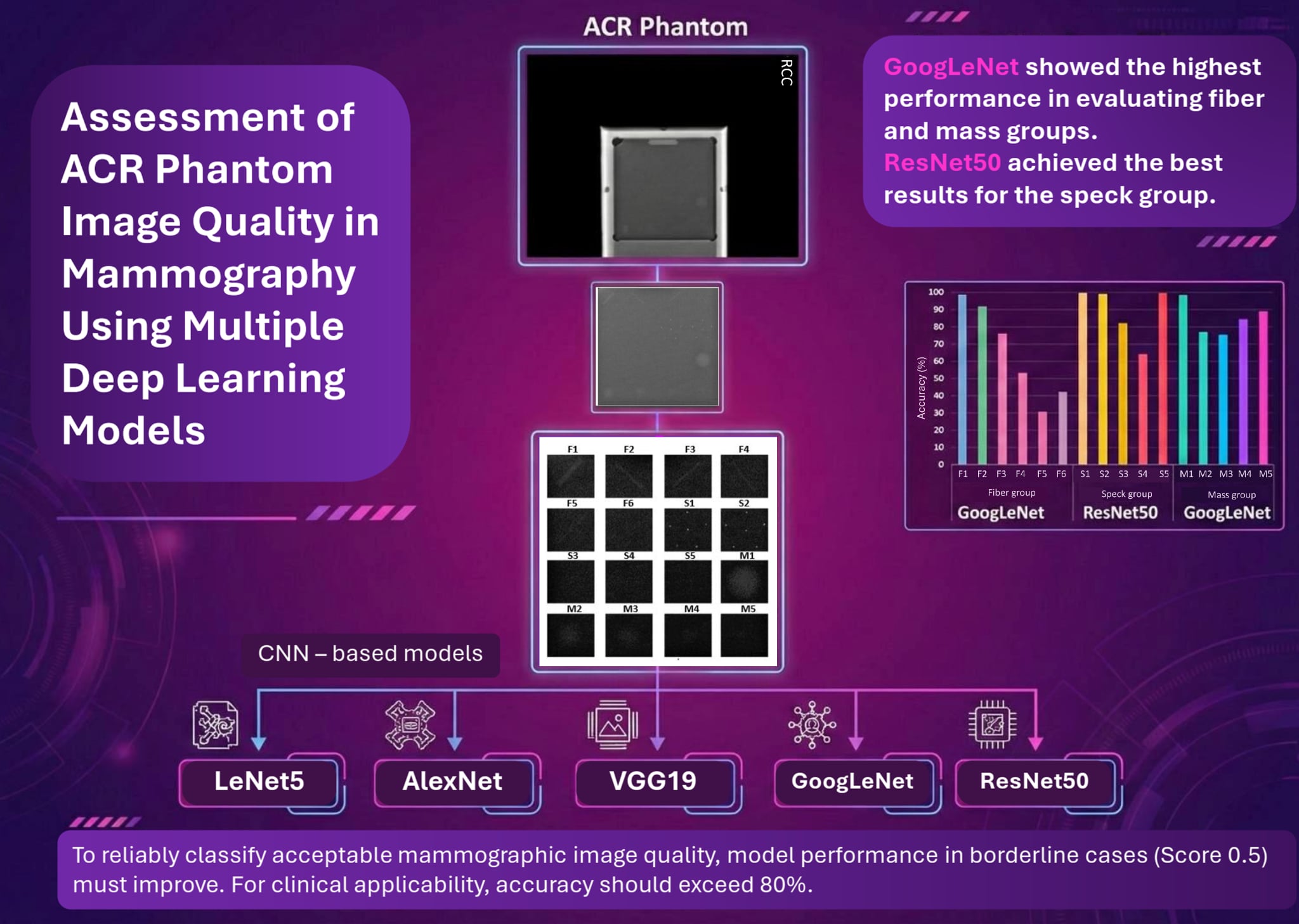
Objectives: This study examined the capability of multiple convolutional neural network (CNN)-based artificial intelligence (AI) models to assess the quality of ACR phantom images and address the limitations of human-based evaluation.
Materials and methods: Five CNN-based models—LeNet5, AlexNet, VGG19, GoogLeNet, and ResNet50—were used to classify 231 ACR phantom images acquired under different exposure settings. Dataset augmentation was performed by adding and removing artificial noise, increasing the dataset to 1,617 images. The dataset was then divided into training (70%), validation (10%), and testing (20%) subsets. Model performance was compared based on phantom image scoring.
Results: GoogLeNet showed the highest performance in evaluating fiber and mass groups, whereas ResNet50 achieved the best results for the speck group. The classification accuracy for scoring 16 object positions in ACR phantom images ranged from 31% to 100%, depending on object size.
Conclusion: To reliably classify acceptable mammographic image quality, model performance in detecting borderline cases (score 0.5) must improve. For clinical applicability, accuracy should exceed 80%.
Article Details

This work is licensed under a Creative Commons Attribution-NonCommercial-NoDerivatives 4.0 International License.
Personal views expressed by the contributors in their articles are not necessarily those of the Journal of Associated Medical Sciences, Faculty of Associated Medical Sciences, Chiang Mai University.
References
Calvocoressi L, Sun A, Kasl S V., Claus EB, Jones BA. Mammography screening of women in their 40s: Impact of changes in screening guidelines. Cancer 2008; 112: 473-80. doi: 10.1002/cncr.23210.
Myers ER, Moorman P, Gierisch JM, Havrilesky LJ, Grimm LJ, Ghate S, et al. Benefits and harms of breast cancer screening: A systematic review. JAMA 2015; 314: 1615-34. doi: 10.1001/jama.2015.13183.
Perry N, Broeders M, de Wolf C, Törnberg S, Holland R, von Karsa L. European guidelines for quality assurance in breast cancer screening and diagnosis. Fourth edition - Summary document. Ann. Oncol. 2008; 19: 614-22. doi: 10.1093/annonc/mdm 481.
Lecun Y, Bottou L, Bengio Y, Haffner P. Gradientbased learning applied to document recognition. Proc. IEEE 1998; 86: 2278-324. doi: 10.1109/5.726791.
Kousalya K, Krishnakumar B, Mohana RS, Karthikeyan N. Comparative analysis of White Blood Cells Classification using Deep Learning Architectures. 2021 2nd International Conference on Smart Electronics and Communication (ICOSEC), 2021; pp. 1220-5. doi: 10.1109/ICOSEC 51865. 2021.9591771.
Jaganathan D, Balsubramaniam S, Sureshkumar V, Dhanasekaran S. Concatenated Modified LeNet Approach for Classifying Pneumonia Images. J Pers Med 2024;14: 328. doi: 10.3390/jpm14030328.
Balasubramaniam S, Velmurugan Y, Jaganathan D, Dhanasekaran S. A modified LeNet CNN for breast cancer diagnosis in ultrasound images. Diagnostics 2023; 13: 2746 doi: 10.3390/diagnostics13172746.
Krizhevsky A, Sutskever I, Hinton GE. ImageNet classification with deep convolutional neural networks. Adv Neural Inf Process Syst. 2012; 25: 1097-105. doi: 10.1145/3065386
Guo C, Chen Y, Li J. Radiographic imaging and diagnosis of spinal bone tumors: AlexNet and ResNet for the classification of tumor malignancy. J Bone Oncol 2024; 48: 100629. doi: 10.1016/j.jbo.2024.100629.
Azhagiri M, Rajesh P. EAN: enhanced AlexNet deep learning model to detect brain tumor using magnetic resonance images. Multimed Tools Appl 2024; 83: 66925-41. doi: 10.1007/s11042-024-18143-w.
Simonyan K, Zisserman A. Very deep convolutional networks for large-scale image recognition. 2015 International Conference on Learning Representations (ICLR), 2015; pp. 1114. https://arxiv.org/abs/1409.1556v6.
Parvin F, Hasan MAM, Ahmed B, Mamun MA, Parvej SMK. Breast cancer histopathological image classification using an ensemble of deep convolutional neural networks. 2023 26th International Conference on Computer and Information Technology (ICCIT), 2023, pp. 1-5. doi.org/10.1109/ICCIT60459.2023.10441273.
Harrison P, Hasan R, Park K. State-of-the-Art of Breast Cancer Diagnosis in Medical Images via Convolutional Neural Networks (CNNs). J Healthc Inform Res. 2023; 7: 387-432. doi: 10.1007/s41666-023-00144-3.
Chen Y, Shao X, Shi K, Rominger A, Caobelli F. AI in breast cancer imaging: An update and future trends. Semin Nucl Med. 2025; 55: 358-70. doi: 10.1053/j.semnuclmed. 2025. 01. 008.
Amanova N, Martin J, Elster C. Explainability for deep learning in mammography image quality assessment. Mach Learn Sci Technol. 2022; 3: 025015. doi: 10.1088/2632-2153/ac7a03.
Szegedy C, Liu W, Jia Y, Sermanet P, Reed S, Anguelov D, Erhan D, Vanhoucke V, Rabinovich A. Going deeper with convolutions. 2015 IEEE Conference on Computer Vision and Pattern Recognition (CVPR), 2015; pp. 1-9. doi: 10.1109/CVPR.2015.7298594.
Teoh JR, Hasikin K, Lai KW, Wu X, Li C. Enhancing early breast cancer diagnosis through automated microcalcification detection using an optimized ensemble deep learning framework. PeerJ Comput Sci 2024;10:e2082. doi: 10.7717/peerj-cs. 2082.
He K, Zhang X, Ren S, Sun J. Deep Residual Learning for Image Recognition. 2016 IEEE Conference on Computer Vision and Pattern Recognition (CVPR), 2016, p. 770–8. doi: 10.1109/CVPR.2016.90.
Zhang Y, Liu YL, Nie K, Zhou J, Chen Z, Chen JH, et al. Deep learning-based automatic diagnosis of breast cancer on MRI using mask R-CNN for detection followed by ResNet50 for classification. Acad Radiol. 2023; 30: S161-71. doi: 10.1016/j.acra.2022.12.038.
Tafavvoghi M, Sildnes A, Rakaee M, Shvetsov N, Bongo LA, Busund L-TR, et al. Deep learningbased classification of breast cancer molecular subtypes from H&E whole-slide images. J Pathol Inform 2025; 16: 100410. doi: 10.1016/j.jpi.2024.100410.
Sundell VM, Mäkelä T, Vitikainen AM, Kaasalainen T. Convolutional neural network -based phantom image scoring for mammography quality control. BMC Med Imaging 2022; 22: 216. doi: 10.1186/s12880-022-00944-w.
Al-antari MA, Al-masni MA, Choi MT, Han SM, Kim TS. A fully integrated computer-aided diagnosis system for digital X-ray mammograms via deep learning detection, segmentation, and classification. Int J Med Inform. 2018; 117: 44-54. doi: 10.1016/j.ijmedinf.2018.06.003.
Ozturk T, Talo M, Yildirim EA, Baloglu UB, Yildirim O, Rajendra Acharya U. Automated detection of COVID-19 cases using deep neural networks with X-ray images. Comput Biol Med. 2020; 121: 103792. doi: 10.1016/j.compbiomed.2020.103792.
Park SS, Ku YM, Seo KJ, Whang IY, Hwang YS, Kim MJ, et al. Devising a deep neural network-based mammography phantom image filtering algorithm using images obtained under mAs and kVp control. Sci Rep. 2023;13: 3545. doi: 10.1038/s41598-023-30780-z.
Ho PS, Hwang YS, Tsai HY. Machine learning framework for automatic image quality evaluation involving a mammographic American College of Radiology phantom. Phys Med. 2022; 102:1-8. doi: 10.1016/j.ejmp.2022.08.004.
Tanaka R, Nozaki S, Goshima F, Shiraishi J. Deep learning versus the human visual system for detecting motion blur in radiography. J Med Imaging. 2022; 9: 015501. doi: 10.1117/1.jmi.9.1.015501.
Song SE, Seo BK, Yie A, Ku BK, Kim HY, Cho KR, et al. Which phantom is better for assessing the image quality in full-field digital mammography?: American College of Radiology accreditation phantom versus digital mammography accreditation phantom. Korean J Radiol. 2012; 13: 776-83. doi: 10.3348/kjr.2012.13.6.776.
Berns EA, Pfeiffer DE, Adent C, Baker JA, Bassett LW, Hendrick FRE, et al. Quality control manual for 2D and digital breast tomosynthesis. Radiologist’s Section, Radiologic Technologist’s Section, Medical Physicist’s Section. American College of Radiology Subcommittee on Quality Assurance in Mammography of the Committee on Mammography Accreditation, American College of Radiology; 2018.
