
Edge-based AI approach for blood vessel segmentation in coronary x-ray angiography
Article Sidebar
Main Article Content
Abstract
Background: An organizational report indicates that heart attacks lead to seventy percent of human fatalities. Heart-related diseases strike people in India who range between the ages of 30-60 years. X-ray coronary angiography functions as the key procedure for detecting these conditions. The manual process of heart vessel segmentation by cardiologists becomes slow and needs significant effort because different professional skill levels affect the consistency of their output results.
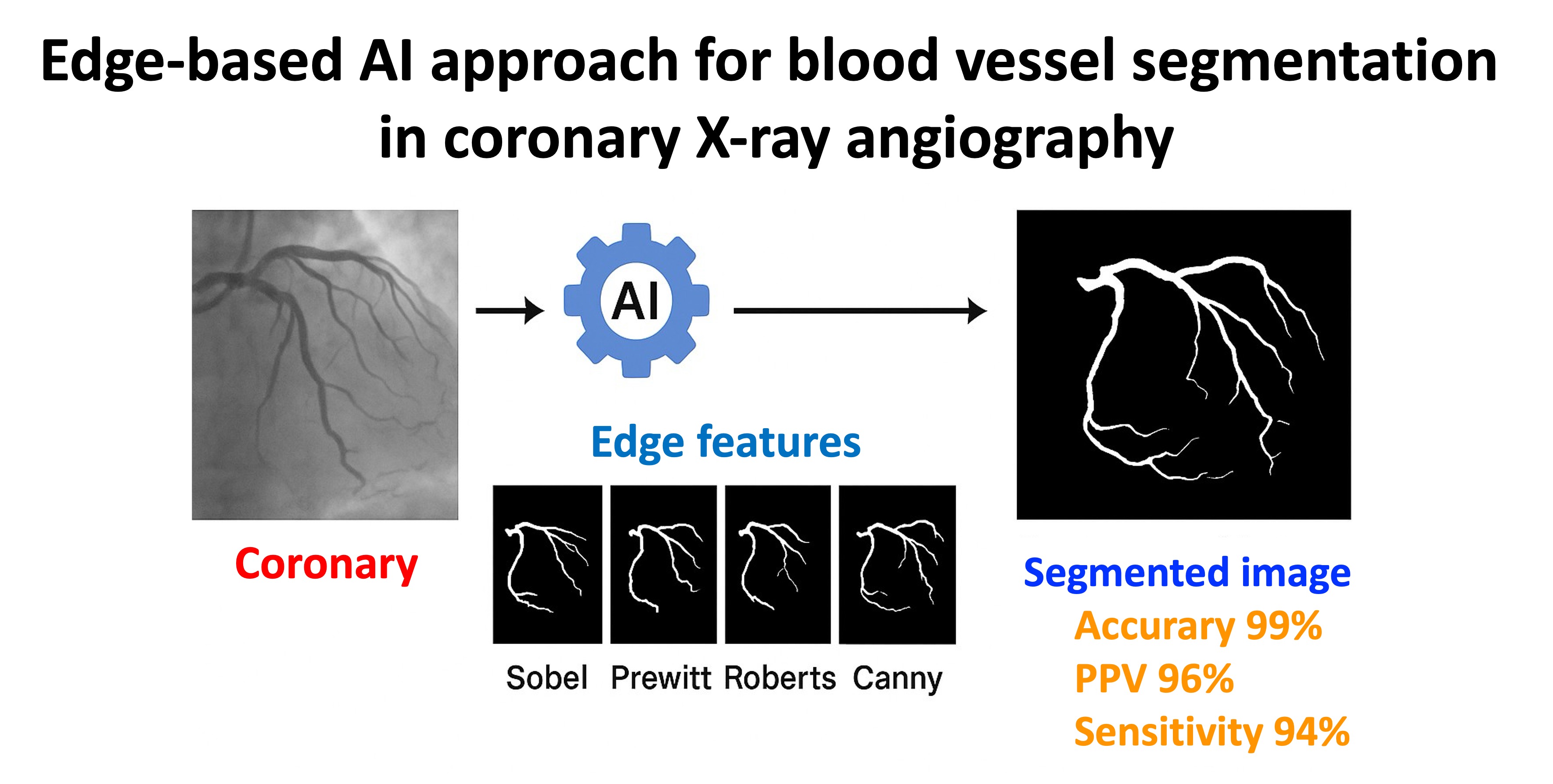
Objective: A study proposes automatic coronary angiography segmentation through artificial intelligence analysis of edge features to accurately detect the main cardiovascular artery system edges.
Materials and methods: The Mendeley public database contained 100 patient images for training purposes and 34 images for validation purposes. The VGG Image Annotator tool served to create binary masks for annotation purposes. The analysis incorporated traditional edge detection methods that included Sobel, Prewitt, and Roberts along with Canny.
Results: The tested model obtained 99% accuracy alongside a positive predictive value (PPV) of 96% and Sensitivity of 94% and Dice Coefficient of 95%. The upcoming research will focus on developing soft computing approaches for detecting stenosis in segmented images.
Conclusion: The method demonstrates better performance metrics that show superior capability to previous techniques implemented in this field. New studies are needed to analyze soft computing techniques to identify vascular structures in coronary angiographic images.
Article Details

This work is licensed under a Creative Commons Attribution-NonCommercial-NoDerivatives 4.0 International License.
Personal views expressed by the contributors in their articles are not necessarily those of the Journal of Associated Medical Sciences, Faculty of Associated Medical Sciences, Chiang Mai University.
References
Di Cesare M, Perel P, Taylor S, Kabudula C, Bixby H, Gaziano TA, et al. The heart of the world. Global Heart. 2024; 19(1): 11. doi: 10.5334/gh.1288.
Abubakar M, Irfan U, Abdelkhalek A, Javed I, Khokhar MI, Shakil F, et al. Comprehensive quality analysis of conventional and novel biomarkers in diagnosing and predicting prognosis of coronary artery disease, acute coronary syndrome, and heart failure, a comprehensive literature review. J Cardiovasc Transl Res. 2024; 17(6): 1258-85. doi: 10.1007/s12265-024-10540-8.
Ren P, He Y, Guo N, Luo N, Li F, Wang Z, et al. A deep learning-based automated algorithm for labeling coronary arteries in computed tomography angiography images. BMC Med Inform Decis Mak. 2023; 23(1): 249. doi: 10.1186/s12911-023-02332-y.
Modric J. Coronary Artery Disease Evidence: Diet, Exercise - eHealthStar [Internet]. eHealthStar - Evidence Based Health Articles. 2016. Available from: https://www.ehealthstar.com/conditions/coronary-heart-disease.
Shukur BS, Mijwil MM. Involving machine learning techniques in heart disease diagnosis: a performance analysis. International J Electr Comput Eng. 2023; 13(2): 2177. doi: 10.11591/ijece.v13i2.pp2177-2185.
Vij R, Arora S. A hybrid evolutionary weighted ensemble of deep transfer learning models for retinal vessel segmentation and diabetic retinopathy detection. Comput Electr Eng. 2024; 115: 109107. doi: 10.1016/j.compeleceng.2024.109107.
Duan X, Sun Y, Wang J. ECA-UNet for coronary artery segmentation and three-dimensional reconstruction. Signal Image Video Process. 2023; 17(3): 783-9. doi:10.1007/s11760-022-02288-y.
Nobre Menezes M, Silva JL, Silva B, Rodrigues T, Guerreiro C, Guedes JP, et al. Coronary X-ray angiography segmentation using Artificial Intelligence: a multi-centric validation study of a deep learning model. Int J Cardiovasc Imaging. 2023; 39(7): 1385-96. doi: 10.1007/s10554-023-02839-5.
Wang G, Zhou P, Gao H, Qin Z, Wang S, Sun J, et al. Coronary vessel segmentation in coronary angiography with a multi-scale U-shaped transformer incorporating boundary aggregation and topology preservation. Phys Med Biol. 2024; 69(2): 025012. doi: 10.1088/1361-6560/ad0b63.
Arefinia F, Aria M, Rabiei R, Hosseini A, Ghaemian A, Roshanpoor A. Non-invasive fractional flow reserve estimation using deep learning on intermediate left anterior descending coronary artery lesion angiography images. Sci Rep. 2024; 14(1): 1818. doi: 10.1038/s41598-024-52360-5.
Zou Z, Li D, Guo H, Yao Y, Yin J, Tao C, et al. Enhancement of structural and functional photoacoustic imaging based on a reference-inputted convolutional neural network. Opt Express. 2025; 33(1): 1260-70. doi: 10.1364/OE.541906.
Rezaee K, Zhu M. Diagnose Alzheimer’s disease and mild cognitive impairment using deep CascadeNet and handcrafted features from EEG signals. Biomed Signal Process Control. 2025; 99: 106895. doi: 10.1016/j.bspc.2024.106895.
Nande SB, Patwardhan SD. Automated Reservoir Characterization of Carbonate Rocks using Deep Learning Image Segmentation Approach. SPE J. 2024; 29(08): 4356-75. doi: 10.2118/219769-PA.
Ren H, Sun Q, Xiao Z, Yu M, Wang S, Yuan L, et al. Heterogeneous feature fusion based machine learning strategy for ECG diagnosis. Expert Syst Appl. 2025: 271: 126714. doi: 10.1016/j.eswa.2025.126714.
Gan H, Zhou R, Ou Y, Wang F, Cheng X, Fu L, et al. A region and category confidence-based multi-task network for carotid ultrasound image segmentation and classification. IEEE J Biomed Health Inform. 2025: online ahead of print. doi: 10.1109/JBHI.2025.3529483.
Mahendiran T, Thanou D, Senouf O, Jamaa Y, Fournier S, De Bruyne B, et al. AngioPy Segmentation: An open-source, user-guided deep learning tool for coronary artery segmentation. Int J Cardiol. 2025; 418: 132598. doi: 10.1016/j.ijcard.2024.132598.
Khan H, Javaid N, Bashir T, Akbar M, Alrajeh N, Aslam S. Heart disease prediction using novel ensemble and blending based cardiovascular disease detection networks: EnsCVDD-Net and BlCVDD-Net. IEEE Access. 2024; 12: 109230-54. doi: 10.1109/ACCESS.2024.3421241.
DeGroat W, Abdelhalim H, Patel K, Mendhe D, Zeeshan S, Ahmed Z. Discovering biomarkers associated and predicting cardiovascular disease with high accuracy using a novel nexus of machine learning techniques for precision medicine. Sci Rep. 2024; 14(1): 1. doi: 10.1038/s41598-023-50600-8.
Qiu Y, Meng J, Li B. Semi-supervised Strong-Teacher Consistency Learning for few-shot cardiac MRI image segmentation. Comput Methods Programs Biomed. 2025; 261: 108613. doi: 10.1016/j.cmpb.2025.108613.
Shi L, Guo H, Liu J. Rapid and automatic hemodynamic assessment: integration of deep learning-based image segmentation, vessel reconstruction, and CFD prediction. Quant Imaging Med Surg. 2025; 15(2): 1358-70. doi: 10.21037/qims-24-1721
Nogueira SA, Luz FA, Camargo TF, Oliveira JC, Campos Neto GC, Carvalhaes FB, et al. Artificial intelligence applied in identifying left ventricular walls in myocardial perfusion scintigraphy images: pilot study. Plos One. 2025; 20(1): e0312257. doi: 10.1371/journal.pone.0312257.
Yu X, Zhu H, Huang B, Hou T, Lu W, Chen N, et al. SCS-SLSP: Hard uncertain pixels mining and utilization for semi-supervised cardiac image segmentation using subjective logic theory and subset prototype generation. Biomed Signal Process Control. 2024; 92: 106145. doi: 10.1016/j.bspc.2024.106145.
Pham TV, Vu TN, Le HM, Pham VT, Tran TT. CapNet: An Automatic Attention-Based with Mixer Model for Cardiovascular Magnetic Resonance Image Segmentation. J Imaging Inform Med. 2025; 38(1): 94-123. doi: 10.1007/s10278-024-01191-x.
Wang G, Zhou M, Ning X, Tiwari P, Zhu H, Yang G, et al. US2Mask: Image-to-mask generation learning via a conditional GAN for cardiac ultrasound image segmentation. Comput Bio Med. 2024; 172: 108282. doi: 10.1016/j.compbiomed.2024.108282.
Danilov V, Klyshnikov K, Kutikhin A, Gerget O, Frangi A, Ovcharenko E. Angiographic dataset for stenosis detection. Mendeley Data. 2021, Version 1. doi:10. 17632/ydrm75xywg.1.
Li H, Xu K. Innovative adaptive edge detection for noisy images using wavelet and Gaussian method. Sci Rep. 2025; 15(1): 5838. doi: 10.1038/s41598-025-86860-9.
Priyanka V, Rama YS, Sravani K, Kavya B. Implementation of Sobel Edge Detection with Image Processing on FPGA. In: 2024 2nd World Conference on Communication & Computing (WCONF) 2024 Jul 12 (pp. 1-5). IEEE. doi: 10.1109/WCONF61366.2024.10692301.
Pritha A, Fathima G. A Detailed Description on Various Techniques of Edge Detection Algorithms. In: Shubham Mahajan, Kapil Joshi, Amit Kant Pandit, Nitish Pathak (Editors). Integrating Metaheuristics in Computer Vision for Real-World Optimization Problems. Crivener Publishing LLC; 2024: pp. 193-205. doi: 10.1002/9781394230952.ch11.
Al Rawahi S. A Comparison of Sobel and Prewitt Edge Detection Operators. EJCS. 2025; 1(1): 49-58. doi: M. Osama and R. Kumar. Journal of Associated Medical Sciences 2025; 58(3): 111-12112110.63496/ejcs.Vol1.Iss1.16.
Zhang R, Jiang G. Exploring a multi-path U-net with probability distribution attention and cascade dilated convolution for precise retinal vessel segmentation in fundus images. Sci Rep. 2025 Apr 18; 15(1): 13428. doi: 10.1038/s41598-025-98021-z
Ylescupidez A, Speake C, Pietropaolo SL, Wilson DM, Steck AK, Sherr JL, et al. OGTT metrics surpass continuous glucose monitoring data for T1D prediction in multiple-autoantibody–positive individuals. J Clin Endocrinol Metab. 2024; 109(1): 57-67. doi: 10.1210/clinem/dgad574.
Challoumis-Κωνσταντίνος Χαλλουμής C. Comparative analysis between capital and liability-Sensitivity Method. OJRE. 2024; 7(2): 63-76. doi: 10.32591/coas.ojre.0702.02063c.
Das N, Das S. Attention-UNet architectures with pretrained backbones for multi-class cardiac MR image segmentation. Curr Prob Cardiol. 2024; 49(1): 102129. doi: 10.1016/j.cpcardiol.2023.102129.
Liu R, Gao S, Zhang H, Wang S, Zhou L, Liu J. MTNet: A combined diagnosis algorithm of vessel segmentation and diabetic retinopathy for retinal images. Plos One. 2022; 17(11): e0278126. doi: 10.1371/journal.pone.0278126.
Cho H, Lee JG, Kang SJ, Kim WJ, Choi SY, Ko J, et al. Angiography-based machine learning for predicting fractional flow reserve in intermediate coronary artery lesions. J Am Heart Assoc. 2019; 8(4): e011685. doi: 10.1161/JAHA.118.011685.
Zhu X, Cheng Z, Wang S, Chen X, Lu G. Coronary angiography image segmentation based on PSPNet. Comput Methods Programs Biomed. 2021; 200:105897. doi: 10.1016/j.cmpb.2020.105897.
Wang L, Liang D, Yin X, Qiu J, Yang Z, Xing J, et al. Coronary artery segmentation in angiographic videos utilizing spatial-temporal information. BMC Med imaging. 2020; 20: 1-0. doi: 10.1186/s12880-020-00509-9.
