
Impact of increasing tube potential and additional filtration on image quality and radiation dose for digital chest radiography
Article Sidebar
Main Article Content
Abstract
Background: Chest radiography is one of the most commonly performed examinations as routine check-ups in radiology departments. Radiographers should be concerned with minimizing patient radiation dose while maintaining high diagnostic image quality.
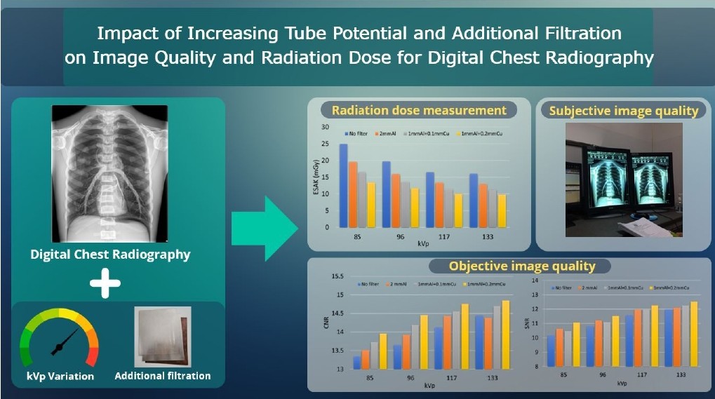
Objective: This study aimed to investigate the effect of increasing tube potential (kV) and adding filtration on image quality and radiation dose for posteroanterior (PA) chest radiography using a digital radiography (DR) system.
Materials and methods: Eighty-five kV with no filter was used as the reference exposure technique. Subsequently, the kV was increased to 96, 117, and 133, and additional filtrations of 2 mm Al, 1 mm Al+0.1 mm Cu, and 1 mm Al+0.2 mm Cu were applied. A total of sixteen images were produced. The entrance surface air kerma (ESAK) was measured and evaluated. Signal-to-noise ratio (SNR) and contrastto-noise ratio (CNR) were accessed for objective image quality. Five independent radiographers assessed a subjective image quality (IQ) score using two alternative forced choices (2AFC).
Results: Increasing kV and adding filtration reduced the ESAK while enhancing the SNR and CNR. However, the IQ score declined relative to the reference image when higher kV and additional filtration were applied except 85 kV. The IQ score indicated that an image acquired at 85 kV with 1 mm Al+0.2 mm Cu showed superior quality compared to the reference image. Notably, the SNR for this image was significantly higher (p<0.05). Additionally, this image resulted in a lower radiation dose (13.44 mGy) compared to the reference image (24.97 mGy). Furthermore, the image quality (IQ) score was higher than the reference images.><0.05). Additionally, this image resulted in a lower radiation dose (13.44 mGy) compared to the reference image (24.97 mGy). Furthermore, the image quality (IQ) score was higher than the reference images.
Conclusion: This study’s findings indicate that using an 85 kV with 1 mm Al+0.2 mm Cu additional filtration for digital PA chest radiography can reduce the radiation dose while improving image quality. However, this study used an anthropomorphic chest phantom; further clinical research is recommended.
Article Details

This work is licensed under a Creative Commons Attribution-NonCommercial-NoDerivatives 4.0 International License.
Personal views expressed by the contributors in their articles are not necessarily those of the Journal of Associated Medical Sciences, Faculty of Associated Medical Sciences, Chiang Mai University.
References
Waheed S, Tahir MJ, Ullah I, Alwalid O, Irshad SG, Asghar MS, et al. The impact of dependence on advanced imaging techniques on the current radiology practice. Ann Med Surg. 2022; 78: 103708. doi.org/ 10.1016/j.amsu.2022.103708
Rehani MM, Nacouzi D. Higher patient doses through X-ray imaging procedures. Phys Med. 2020; 79: 80-6. doi.org/10.1016/j.ejmp.2020.10.017
Smith-Bindman R, Miglioretti DL, Larson EB. Rising use of diagnostic medical imaging in a large integrated health system. Health Aff (Millwood). 2008; 27(6): 1491-502. doi.org/10.1377/hlthaff.27.6.1491
Schaefer-Prokop C, Neitzel U, Venema HW, Uffmann M, Prokop M. Digital chest radiography: an update on modern technology, dose containment and control of image quality. Eur Radiol. 2008; 18(9): 1818-30. doi. org/10.1007/s00330-008-0948-3
Atiyyah TAE-R, Nasr MSN, Ahmed TS, Mostafa MMSA. Cumulative radiation exposure from diagnostic imaging in Zagazig University Pediatric Intensive Care and Chest Units. Egypt J Hosp Med. 2020; 81(2): 1520-4. doi.org/10.21608/ejhm.2020.115566
Little MP, Wakeford R, Tawn EJ, Bouffler SD, Berrington de Gonzalez A. Risks associated with low doses and low dose rates of ionizing radiation: why linearity may be (almost) the best we can do. Radiology. 2009; 251(1): 6-12. doi.org/10.1148/radiol.2511081686
The 2007 Recommendations of the International Commission on Radiological Protection. ICRP publication 103. Ann ICRP. 2007; 37(2-4): 1-332. doi. org/10.1016/j.icrp.2007.10.003
Martin C. Optimisation in general radiography. Biomed Imaging Interv J. 2007; 3(2): e18. doi.org/10. 2349/biij.3.2.e18
Smans K, Struelens L, Smet M, Bosmans H, Vanhavere F. Cu filtration for dose reduction in neonatal chest imaging. Radiat Prot Dosim. 2010; 139(1-3): 281-6. doi.org/10.1093/rpd/ncq061
Papadakis AE, Giannakaki V, Hatzidaki E, Damilakis J. The effect of added filtration on radiation dose and image quality in digital radiography of newborns. Pediatr Radiol. 2023; 53(10): 2060-8. doi.org/10. 1007/s00247-023-05698-3
Bushong SC. Radiologic science for technologists: Physics, biology, and protection. 10th Ed. USA: Elsevier Health Sciences; 2013. https://books.google.co.th/ books?id=I7LSCQAAQBAJ
Carroll QB, Fuchs AW. Fuchs’s radiographic exposure, processing, and quality control: Charles C. Thomas; 1998. https://books.google.co.th/books?id=EcZpA AAAMAAJ
Ching W, Robinson J, McEntee M. Patient-based radiographic exposure factor selection: a systematic review. J Med Radiat Sci. 2014; 61(3): 176-90. doi. org/10.1002/jmrs.66
Pernicka F, McLean I. Dosimetry in diagnostic radiology: an international code of practice: International Atomic Energy Agency Vienna, Austria; 2007.
Alzyoud K, Hogg P, Snaith B, Flintham K, England A. Impact of body part thickness on AP pelvis radiographic image quality and effective dose. Radiography. 2019; 25(1): e11-e7. doi.org/10.1016/j.radi.2018.09.001
Nocetti D, Villalobos K, Marín N, Monardes M, Tapia B, Toledo MI, et al. Radiation dose reduction and image quality evaluation for lateral lumbar spine projection. Heliyon. 2023; 9(9): e19509. doi.org/ 10.1016/j.heliyon.2023.e19509
Tugwell J, Everton C, Kingma A, Oomkens DM, Pereira GA, Pimentinha DB, et al. Increasing source to image distance for AP pelvis imaging – Impact on radiation dose and image quality. Radiography. 2014; 20(4): 351-5. doi.org/10.1016/j.radi.2014.05.012
European Commission, Directorate-General for Research Innovation, Carmichael J, Moores B, Maccia C. European guidelines on quality criteria for diagnostic radiographic images: Publications Office; 1996. https://op.europa.eu/en/publication-detail/-/publication/ d59ccc60-97ed-4ce8-b396-3d2d42b284be
Koo TK, Li MY. A Guideline of selecting and reporting intraclass correlation coefficients for reliability research. J Chiropr Med. 2016; 15(2): 155-63. doi.org/10.1016 /j.jcm.2016.02.012
Stewart C, Raja H, Torrance E, Funk L. In Vivo randomized controlled study of the bone response of all-suture anchors and biocomposite anchors. Orthop J Sports Med. 2020; 8: 232596712091496. doi.org/10.1177/2325967120914965
Han X. On statistical measures for data quality evaluation. J Geogr Inf Syst. 2020; 12: 178-87. doi. org/10.4236/jgis.2020.123011
Fauber TL. Radiographic imaging & exposure. 5th Ed. St. Louis, Mo.: Elsevier; 2017.
Mifsud K, Portelli JL, Zarb F, Couto JG. Evaluating the use of higher kVp and copper filtration as a dose optimisation tool in digital planar radiography. Radiography. 2022; 28(3): 586-92. doi.org/10.1016/j. radi.2022.04.002
Jang JS, Yang HJ, Koo HJ, Kim SH, Park CR, Yoon SH, et al. Image quality assessment with dose reduction using high kVp and additional filtration for abdominal digital radiography. Physica Medica. 2018; 50: 46-51. doi.org/10.1016/j.ejmp.2018.05.007
Kawashima H, Ichikawa K, Nagasou D, Hattori M. X-ray dose reduction using additional copper filtration for abdominal digital radiography: Evaluation using signal difference-to-noise ratio. Physica Medica. 2017; 34: 65-71. doi.org/10.1016/j.ejmp.2017.01.015
Ekpo EU, Hoban AC, McEntee MF. Optimisation of direct digital chest radiography using Cu filtration. Radiography. 2014; 20(4): 346-50. doi.org/10.1016/j. radi.2014.07.001
Huda W, Abrahams RB. Radiographic techniques, contrast, and noise in X-Ray imaging. Am J Roentgenol. 2015; 204(2): W126-W31. doi.org/10.2214/ajr.14.13116
