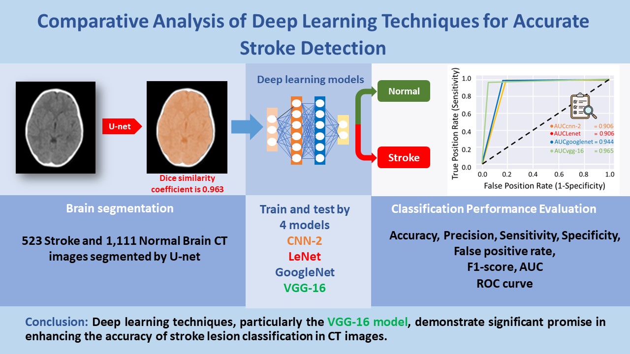
Comparative analysis of deep learning techniques for accurate stroke detection
Main Article Content
Abstract
Background: The traditional diagnosis of strokes through computed tomography (CT) heavily relies on radiologists’ expertise for accurate interpretation. However, the increasing demand for this critical task exceeds the available radiologist workforce, necessitating innovative solutions. This research addresses this challenge by introducing deep learning techniques to enhance the initial screening of stroke cases, thereby augmenting radiologists’ diagnostic capabilities.
Objective: This study aims to compare four techniques for classifying stroke lesions in CT images.
Materials and methods: Four distinct models-CNN-2-Model, LeNet, GoogleNet, and VGG-16-were trained using a dataset comprising 1,636 CT images, including 1,111 normal brain images and 525 stroke images. Seventy percent of the images were used to train the most effective deep learning model, and subsequently, these images were utilized to evaluate the performance of each model. The evaluation involved assessing accuracy, precision, sensitivity, specificity, F1 score, false positive rate, and AUC.
Results: The evaluation process included a comprehensive statistical analysis of the models’ prediction results. The findings revealed that VGG-16 emerged as the top-performing deep learning model, achieving an impressive accuracy of 0.969, precision of 0.952, sensitivity of 0.952, specificity of 0.978, F1 score of 0.952, false positive rate of 0.022, and AUC of 0.965.
Conclusion: In conclusion, deep learning techniques, particularly the VGG-16 model, demonstrate significant promise in enhancing the accuracy of stroke lesion classification in CT images. These findings underscore the potential of leveraging advanced technologies to address the growing challenges in stroke diagnosis and pave the way for more efficient and accessible healthcare solutions.
Article Details

This work is licensed under a Creative Commons Attribution-NonCommercial-NoDerivatives 4.0 International License.
Personal views expressed by the contributors in their articles are not necessarily those of the Journal of Associated Medical Sciences, Faculty of Associated Medical Sciences, Chiang Mai University.
References
Amarenco P, Bogousslavsky J, Caplan LR, Donnan GA, Hennerici MG. Classification of Stroke Subtypes. Cerebrovasc Dis. 2009; 27 (5): 493-501. doi.org/10.1159/000210432
World Health Organization (WHO). World stroke day 2022 [cited 2022 November 17]. Available from: https://www.who.int/srilanka/news/detail/29-10-2022-world-stroke-day-2022
The Ministry of Public Health of Thailand, Public Health Statistics A.D. 2022. [cited 2022 May 14]. Available from: https://spd.moph.go.th/wp-content/uploads/2023/11/Hstatistic65.pdf
González RG. Current State of Acute Stroke Imaging. Stroke. 2013; 44(11): 3260-4. doi.org/10.1161/STROKEAHA.113.003229
Birenbaum D, Bancroft LW, Felsberg GJ. Imaging in Acute Stroke. West J Emerg Med. 2011; 12(1): 67-76. https://www.ncbi.nlm.nih.gov/pmc/articles/PMC3088377/
Raghavendra U, Gudigar A, Paul Aritra, Goutham TS, Inamdar MA, Hegde A, et al. Brain tumor detection and screening using artificial intelligence techniques: Current trends and future perspectives. Comput Biol Med. 2023; 163: 107063. doi.org/10.1016/j.compbiomed.2023.107063
Yousaf F, Iqbal S, Fatima N, Kousar T, Rahim MSM. Multi-class disease detection using deep learning and human brain medical imaging. Biomed Signal Process Control. 2023; 85: 104875. doi.org/10.1016/j.bspc.2023.104875
Ammar M, Lamria MA, Mahmoudib S, Laidia A. Deep Learning Models for Intracranial Hemorrhage Recognition: A comparative study. Procedia Comput Sci. 2022; 196: 418-25. doi.org/10.1016/j.procs.2021.12.031
Worachotsueptrakun C. A comparison of 3D Convolutional neural network for brain stroke classification with CT scan images. Chulalongkorn University theses and dissertations. 2022. https://digital.car.chula.ac.th/chulaetd/6664
Vamsi B, Bhattacharyya D, Midhunchakkravarthy D, Kim JY. Early Detection of Hemorrhagic Stroke Using a Lightweight Deep Learning Neural Network Model. Trait du signal. 2021; 38(6)1727-36. doi.org/10.18280/ts.380616
Tasnia N. brain-stroke-prediction-ct-scan-image-dataset. [cited 2022 May 1]. Available from: https://www. kaggle.com/datasets/noshintasnia/brain-strokeprediction-ct-scan-image-dataset
Vbookshelf. Brain CT Images with Intracranial Hemorrhage Masks. [cited 2022 May 1]. Available from: https://www.kaggle.com/datasets/vbookshelf/computed-tomography-ct-images
Pandey N. Lung segmentation from Chest X-Ray dataset. Kaggle Inc [cited 2022 April 28]. Available from: https://www.kaggle.com/code/nikhilpandey360/lung-segmentation-from-chest-x-ray-dataset/notebook
Chen YT, Chen YL, Chen YY, Huang YT, Wong HF, Yan JL, Wang JJ. Deep Learning–Based Brain Computed Tomography Image Classification with Hyperparameter Optimization through Transfer Learning for Stroke. Diagnostics 2022; 12, 807. doi.org/10.3390/diagnostics12040807
Ozaltin O, Coskun O, Yeniay O, Subasi A. A Deep Learning Approach for Detecting Stroke from Brain CT Images Using OzNet. Bioeng. 2022; 9(12): 783. doi.org/10.3390/bioengineering9120783


