
Analyzing DNA barcoding and identifying toxins caused by neurotoxic mushroom poisoning using liquid chromatography tandem mass spectrometry
Article Sidebar
Main Article Content
Abstract
Background: Neurotoxic mushroom poisoning often exhibits rapid symptom onset, typically attributed to compounds such as Ibotenic acid, which affect the central nervous system. This study addresses a new case of mushroom-related food poisoning in southern Thailand.
Objective: The objectives are to determine the presence of ibotenic acid in cases of mushroom-related food poisoning utilizing liquid chromatography-tandem mass spectrometry (LC-MS/MS) and to identify toxic Amanita species implicated in these cases.
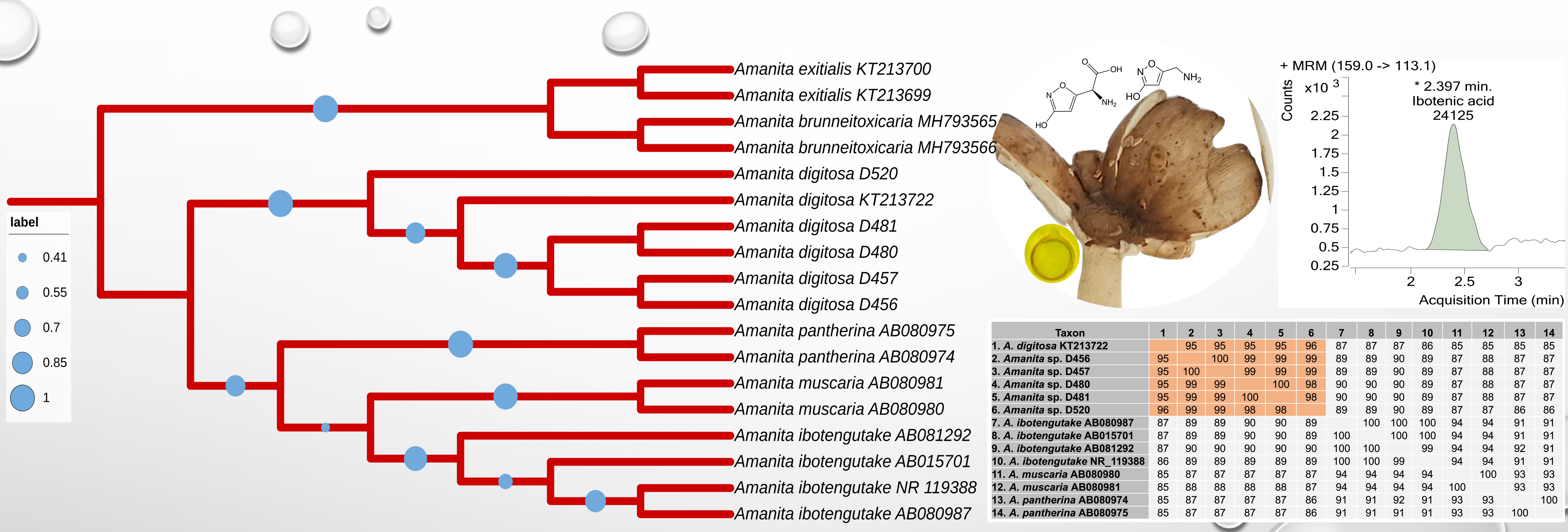
Materials and methods: Remnant mushroom samples obtained from three clinically reported cases were used. Nucleotide similarity was compared against the rRNA/ ITS databases using NCBI BLAST search. Phylogenetic analyses were conducted using maximum likelihood (ML) and FastTree approaches. LC-MS/MS was employed to separate of Ibotenic acid, determine its molecular weight and perform precursor ion fragmentation.
Results: Analysis of the rRNA/ITS databases revealed a high nucleotide similarity between suspected mushroom samples and Amanita digitosa. Detailed phylogenetic analysis confirmed that mushroom samples from the three poisoning cases clustered with A. digitosa. LC-MS/MS analysis showed the presence of ibotenic acid, with precursor ion (m/z 159) and product ion (m/z 113.1) as the major toxic substances. Clinically, patients poisoned by ibotenic acid-containing mushrooms exhibited a short latent period with symptoms of nausea, vomiting, vertigo, delirium, confusion, and fatigue.
Conclusion: The genus Amanita comprises both edible and inedible species that produce several lethal toxins. The report of ibotenic acid in A. digitosa is a novel finding, valuable for food safety monitoring and healthcare decision-maker. This is especially notable due to the accuracy and rapidity of the analytical process.
Article Details

This work is licensed under a Creative Commons Attribution-NonCommercial-NoDerivatives 4.0 International License.
Personal views expressed by the contributors in their articles are not necessarily those of the Journal of Associated Medical Sciences, Faculty of Associated Medical Sciences, Chiang Mai University.
References
Ratnasingham S, Hebert PDN. bold: The Barcode of Life Data System (http://www.barcodinglife.org). Mol Ecol Notes. 2007; 7(3): 355-64. doi.org/10.1111/j.1471-8286.2007.01678.x.
Parnmen S, Sikaphan S, Leudang S, Boonpratuang T, Rangsiruji A, Naksuwankul K. Molecular identification of poisonous mushrooms using nuclear ITS region and peptide toxins: a retrospective study on fatal cases in Thailand. J Toxicol Sci. 2016; 41(1): 65-76.
Parnmen S, Nooron N, Leudang S, Sikaphan S, Polputpisatkul D, Rangsiruji A. Phylogenetic evidence revealed Cantharocybe virosa (Agaricales, Hygrophoraceae) as a new clinical record for gastrointestinal mushroom poisoning in Thailand. Toxicol Res. 2020; 36(3): 239-48. doi.org/10.1007/s43188-019-00024-2.
Ramchiun S, Sikaphan S, Leudang S, Polputpisatkul D, Nantachaiphong N, Khaentaw T, et al. Molecular characterization and liquid chromatography-mass spectrometric multiple reaction monitoring-based detection in case of suspected phalloides syndrome poisoning. J Assoc Med Sci. 2019; 52(1): 48-55. doi: 10.14456/jams.2019.9.
Leudang S, Sikaphan S, Parnmen S, Nantachaiphong N, Polputpisatkul D, Ramchiun S, et al. DNA-based identification of gastrointestinal irritant mushrooms in the genus Chlorophyllum: A food poisoning case in Thailand. J Heal Res. 2017; 31(1): 41-9. doi: 10.14456/jhr.2017.6.
Parnmen S, Nooron N, Leudang S, Sikaphan S, Polputpisatkul D, Pringsulaka O, et al. Foodborne illness caused by muscarine-containing mushrooms and identification of mushroom remnants using phylogenetics and LC-MS/MS. Food Control. 2021; 128: 108182. doi.org/10.1016/j.foodcont.2021.108182.
Nooron N, Parnmen S, Chonnakijkul P, Sikaphan S, Chankunasuka R, Phatsarapongkul S, et al. Use of nuclear ITS region as DNA barcode marker for the species identification of mushroom in the genus Macrocybe causing foodborne illness. Thai J Toxicol. 2023; 38(1): 55-67. Available from: https://li01.tcithaijo.org/index.php/ThaiJToxicol/article/view/258482.
Xu J. Fungal DNA barcoding. Genome. 2016; 59(11): 913-32. doi: 10.1139/gen-2016-0046.
Schoch CL, Seifert KA, Huhndorf S, Robert V, Spouge JL, Levesque CA, et al. Nuclear ribosomal internal transcribed spacer (ITS) region as a universal DNA barcode marker for Fungi. Proc Natl Acad Sci USA. 2012; 109(16): 6241-6. doi: 10.1073/pnas.1117018109.
Nooron N, Parnmen S, Sikaphana S, Leudang S, Uttawichai C, Polputpisatkul D. The situation of mushrooms food poisoning in Thailand: symptoms and common species list. Thai J Toxicol. 2020; 35(2): 58-69. Available from: https://li01.tci-thaijo.org/index.php/ThaiJToxicol/article/view/248088.
White J, Weinstein SA, De Haro L, Bédry R, Schaper A, Rumack BH, et al. Mushroom poisoning: A proposed new clinical classification. Toxicon. 2019; 157: 53-65. doi: 10.1016/j.toxicon.2018.11.007.
Li GJ, Hyde KD, Zhao RL, Hongsanan S, Abdel-Aziz FA, Abdel-Wahab MA, et al. Fungal diversity notes 253--366: taxonomic and phylogenetic contributions to fungal taxa. Fungal Divers. 2016; 1-237. doi.org/10.1007/s13225-016-0366-9.
Trakulsrichai S, Jeeratheepatanont P, Sriapha C, Tongpoo A, Wananukul W. Myotoxic mushroom poisoning in Thailand: clinical characteristics and outcomes. Int J Gen Med. 2020; 13: 1139-46. doi: 10.2147/IJGM.S271914.
He M-Q, Wang M-Q, Chen Z-H, Deng W-Q, Li T-H, Vizzini A, et al. Potential benefits and harms: a review of poisonous mushrooms in the world. Fungal Biol Rev. 2022; 42: 56-68.
Duffy TJ. Toxic Fungi of Western North America. Vol. 2008, Group. MykoWeb; 2008. 166 p. Available from: www.mykoweb.com
Gardes M, Bruns TD. ITS primers with enhanced specificity for basidiomycetes--application to the identification of mycorrhizae and rusts. Mol Ecol. 1993; 2(2): 113-8. doi.org/10.1111/j.1365-294X.1993.tb00005.x.
White TJ, Bruns TD, Lee SB, Taylor JW. Amplification and direct sequencing of fungal ribosomal RNA genes for phylogenetics. In: Innis, M.A., Gelfand, D.H., Sninsky, J.J. & White T., editor. PCR Protocols: a guide to methods and applications. New York: Academic Press; 1990. p. 315-22.
Altschul SF, Gish W, Miller W, Myers EW, Lipman DJ. Basic local alignment search tool. J Mol Biol. 1990; 215(3): 403-10. doi.org/10.1016/S0022-2836(05)80360-2.
Department of Disease Control. Mushroom poisoning. National Disease Surveillance (Report 506). 2023. Available from: http://doe.moph.go.th/surdata/index.php
Saingam D, Assanangkornchai S, Geater AF, Balthip Q. Pattern and consequences of krathom (Mitragyna speciosa Korth.) use among male villagers in southern Thailand: a qualitative study. Int J Drug Policy. 2013; 24(4): 351-8. doi: 10.1016/j.drugpo.2012.09.004.
Oda T, Yamazaki T, Tanaka C, Terashita T, Taniguchi N, Tsuda M. Amanita ibotengutake sp. nov., a poisonous fungus from Japan. Mycol Prog. 2002; 1(4): 355-65. doi.org/10.1007/s11557-006-0032-9.
Tsujikawa K, Kuwayama K, Miyaguchi H, Kanamori T, Iwata Y, Inoue H, et al. Determination of muscimol and ibotenic acid in Amanita mushrooms by highperformance liquid chromatography and liquid chromatography-tandem mass spectrometry. J Chromatogr B, Anal Technol Biomed life Sci. 2007; 852(1-2): 430-5. doi: 10.1016/j.jchromb.2007.01.046.
Bresinsky A, Besl H. A colour atlas of poisonous fungi: A handbook for pharmacists, doctors, and biologists. London: Wolfe Publishing Ltd; 1990. P.1-295.
Hiroshima Y, Nakae H, Gommori K.Amanita ibotengutake intoxication treated with plasma exchange. Ther Apher Dial. 2010; 14(5): 483-4. doi: 10.1111/j.1744-9987.2010.00862.x.
