
Evaluation of the offset couch parameter between kilovoltage on-board imaging and cone-beam computed tomography in patients with prostate cancer
Article Sidebar
Main Article Content
Abstract
Background: External Beam Radiation Therapy (EBRT) is a curative therapy technique for prostate cancer. Since the prostate is unstable and surrounded by the bladder and rectum, precision of the target location is critical. Image Guided Radiation Therapy (IGRT) can improve treatment precision. The bladder and rectum may alter volume during IGRT, shifting the prostate’s position and resulting in missed target volume doses and extra organs at risk (OARs) doses.
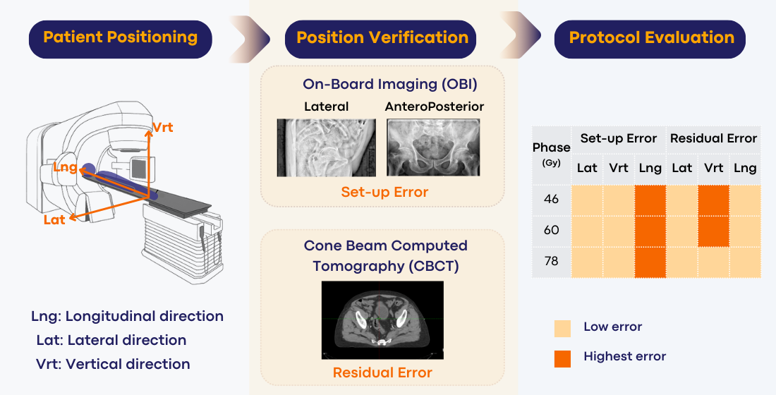
Objective: To assess setup error and residual error during patient positioning, as well as the current IGRT protocol efficiency, in prostate cancer patients while recommending a planning target volume (PTV) margin.
Materials and methods: The offset couch parameter of on-board imaging (OBI) and cone-beam computed tomography (CBCT) was computed to determine the error distribution, magnitude, and error difference between treatment phases. The systematic and random errors were calculated using the van Herk equation to determine the planning target volume (PTV) margin.
Results: The setup error was -0.86 to 0.25 mm, and the residual error was -0.15 to 0.32 mm. The couch displacement percentage for OBI was 29.44% to 58.89%, and for CBCT was 8.10% to 34.12%. The systematic error was 1.65 to 3.21. The random error was 1.78 to 3.29. The setup error was greatest in the longitudinal (Lng) direction, residual error was greatest in the vertical (Vrt) direction, and systematic and random error were greatest in the Vrt and lateral (Lat) direction, respectively. The PTV margin was greatest in the Vrt direction, while the Lng direction was the narrowest margin for every treatment phase.
Conclusion: The highest setup error occurs in the Lng direction for all treatment phases. For the 46 Gy and 60 Gy phases, the highest residual error is in the Vrt direction. However, in the 78 Gy phase, the error is relatively close to 0.01mm in every direction. The current IGRT protocol is effective in detecting setup and residual errors. The 78 Gy phase has the greatest PTV margin, whereas the 46 Gy phase shows the narrowest margins in all directions.
Article Details

This work is licensed under a Creative Commons Attribution-NonCommercial-NoDerivatives 4.0 International License.
Personal views expressed by the contributors in their articles are not necessarily those of the Journal of Associated Medical Sciences, Faculty of Associated Medical Sciences, Chiang Mai University.
References
Naravejsakul k, Pothisa T, Saenrak N. Incidence of Prostate Cancer in Physical Checkup Population with Rising of Serum Prostatic Specific Antigen. Vajira Med J. 2022; 66(5): 361-8. doi: 10.14456/vmj.2022.37.
Somboon S, Malila W, Tamon S, Nueangwong W, Yeenang N, Rueansri J. Evaluation of optimal kilovoltagecone beam technique on image quality, registration accuracy, time of imaging and relative dose for head radiotherapy: A phantom study. J Assoc Med Sci. 2022; 55(2): 10-5. doi: 10.12982/JAMS.2022.011.
Huang K, Palma D, Scott D, McGregor D, Yartsev S, Louie A, et al. Inter- and Intrafraction Uncertainty in Prostate Bed Image-Guided Radiotherapy. Int. J. Radiat. Oncol Biol Phys. 2012; 84: 402-7. doi: 10.1016/j.ijrobp.2011.12.035.
Ariyaratne H, Chesham H, Pettingell J, Alonzi R. Imageguided radiotherapy for prostate cancer with cone beam CT: dosimetric effects of imaging frequency and PTV margin. Radiother Oncol. 2016; 121(1): 103-8. doi: 10.1016/j.radonc.2016.07.018.
Ghadjar P, Fiorino C, Munck Af Rosenschöld P, Pinkawa M, Zilli T, van der Heide UA. ESTRO ACROP consensus guideline on the use of image guided radiation therapy for localized prostate cancer. Radiother Oncol. 2019; 141: 5-13. doi: 10.1016/j.radonc.2019.08.027.
van Herk M. Errors and margins in radiotherapy. Semin Radiat Oncol. 2004; 14(1): 52-64. doi: 10.1053/j.semradonc.2003.10.003.
Hashido T, Nakasone S, Fukao M, Ota S, Inoue S. Comparison between manual and automatic image registration in image-guided radiation therapy using megavoltage cone-beam computed tomography with an imaging beam line for prostate cancer. Radiol Phys Technol. 2018; 11(4): 392-405. doi: 10.1007/s12194-018-0476-z.
Hurkmans CW, Remeijer P, Lebesque JV, Mijnheer BJ. Set-up verification using portal imaging; review of current clinical practice. Radiother Oncol. 2001; 58(2): 105-20. doi: 10.1016/s0167-8140(00)00260-7.
Mayyas E, Chetty IJ, Lu M, Stricker H, Pradhan D, Movsas B, et al. Evaluation of multiple image-based modalities for image-guided radiation therapy (IGRT) of prostate carcinoma: a prospective study. Med Phys. 2013; 40(4): 041707. doi: 10.1118/1.4794502.
Ghilezan MJ, Jaffray DA, Siewerdsen JH, Van Herk M, Shetty A, Sharpe MB, et al. Prostate gland motion assessed with cine-magnetic resonance imaging (cine-MRI). Int J Radiat Oncol Biol Phys. 2005; 62(2): 406-17. doi: 10.1016/j.ijrobp.2003.10.017.
McNair HA, Hansen VN, Parker CC, Evans PM, Norman A, Miles E, et al. A comparison of the use of bony anatomy and internal markers for offline verification and an evaluation of the potential benefit of online and offline verification protocols for prostate radiotherapy. Int J Radiat Oncol Biol Phys. 2008; 71(1): 41- 50. doi: 10.1016/j.ijrobp.2007.09.002.
Zucca S, Carau B, Solla I, Garibaldi E, Farace P, Lay G, et al. Prostate image-guided radiotherapy by megavolt cone-beam CT. Strahlenther Onkol. 2011; 187(8): 473-8. doi: 10.1007/s00066-011-2241-7.
Poulsen PR, Muren LP, Høyer M. Residual set-up errors and margins in on-line image-guided prostate localization in radiotherapy. Radiother Oncol. 2007; 85(2): 201-6. doi: 10.1016/j.radonc.2007.08.006.
van Herk M, Bruce A, Kroes AP, Shouman T, Touw A, Lebesque JV. Quantification of organ motion during conformal radiotherapy of the prostate by three dimensional image registration. Int J Radiat Oncol Biol Phys. 1995; 33(5): 1311-20. doi: 10.1016/0360-3016(95)00116-6.
Ost P, De Meerleer G, De Gersem W, Impens A, De Neve W. Analysis of prostate bed motion using daily cone-beam computed tomography during postprostatectomy radiotherapy. Int J Radiat Oncol Biol Phys. 2011; 79(1): 188-94. doi: 10.1016/j.ijrobp.2009.10.029.
Nakamura N, Shikama N, Takahashi O, Ito M, Hashimoto M, Uematsu M, et al. Variability in bladder volumes of full bladders in definitive radiotherapy for cases of localized prostate cancer. Strahlenther Onkol. 2010; 186(11): 637-42. doi: 10.1007/s00066-010-2105-6.
Schallenkamp JM, Herman MG, Kruse JJ, Pisansky TM. Prostate position relative to pelvic bony anatomy based on intraprostatic gold markers and electronic portal imaging. Int J Radiat Oncol Biol Phys. 2005; 63(3): 800-11. doi: 10.1016/j.ijrobp.2005.02.022.
Millender LE, Aubin M, Pouliot J, Shinohara K, Roach M, 3rd. Daily electronic portal imaging for morbidly obese men undergoing radiotherapy for localized prostate cancer. Int J Radiat Oncol Biol Phys. 2004; 59(1): 6-10. doi: 10.1016/j.ijrobp.2003.12.027.
Moseley DJ, White EA, Wiltshire KL, Rosewall T, Sharpe MB, Siewerdsen JH, et al. Comparison of localization performance with implanted fiducial markers and cone-beam computed tomography for on-line image-guided radiotherapy of the prostate. Int J Radiat Oncol Biol Phys. 2007; 67(3): 942-53. doi: 10.1016/j.ijrobp.2006.10.039.
Bylund KC, Bayouth JE, Smith MC, Hass AC, Bhatia SK, Buatti JM. Analysis of interfraction prostate motion using megavoltage cone beam computed tomography. Int J Radiat Oncol Biol Phys. 2008; 72(3): 949-56. doi: 10.1016/j.ijrobp.2008.07.002.
van Herk M, Remeijer P, Rasch C, Lebesque JV. The probability of correct target dosage: dose-population histograms for deriving treatment margins in radiotherapy. Int J Radiat Oncol Biol Phys. 2000; 47(4): 1121-35. doi: 10.1016/s0360-3016(00)00518-6.
Ding GX, Coffey CW. Radiation dose from kilovoltage cone beam computed tomography in an imageguided radiotherapy procedure. Int J Radiat Oncol Biol Phys. 2009; 73(2): 610-7. doi: 10.1016/j.ijrobp. 2008.10.006.
Wang G, Wang WL, Liu YQ, Dong HM, Hu YX. Positioning error and expanding margins of planning target volume with kilovoltage cone beam computed tomography for prostate cancer radiotherapy. Onco Targets Ther. 2018; 11: 1981-8. doi: 10.2147/ott.S152915.
