
Two-stage method for hepatocellular carcinoma screening in B-mode ultrasound images
Article Sidebar
Main Article Content
Abstract
Background: Hepatocellular carcinoma (HCC) is a significant global health concern that requires early detection for effective treatment.
Objectives: The objective of this study was to develop a system for screening HCC in B-mode ultrasound images.
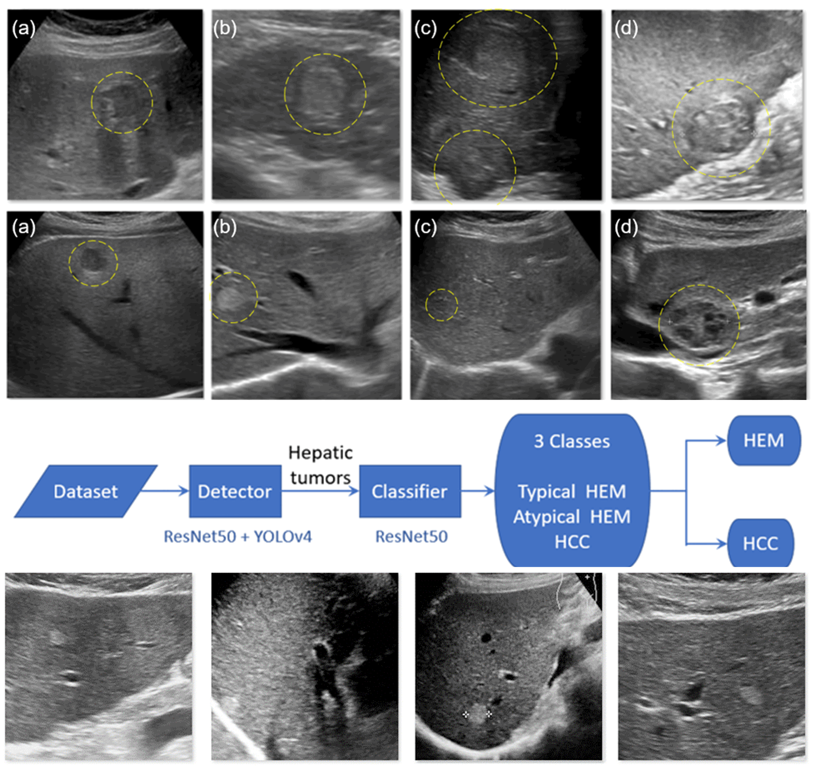
Materials and methods: The dataset consisted of 1665 hemangioma (HEM) images, including 961 typical HEM, 704 atypical HEM, and 543 HCC images. Four YOLOv4 models were trained: one for HCC detection, one for the conventional two-class detection of HEM and HCC, one to detect typical HEM and suspicious lesions, and the last one was our two-stage model consisting of a detector and classifier. In the first stage, a YOLOv4-based detector with ResNet-50 as the backbone was used to identify focal liver lesions. The second stage utilized ResNet-50 as a classifier to classify the lesions into HCC, atypical HEM, or typical HEM. Differentiating between HCC and atypical HEM is not necessary, as both require further investigation with CT or MR imaging.
Results: The evaluation of the developed HCC screening system using ten-fold cross-validation showed that grouping HCC and atypical HEM together significantly increased precision from 0.74 to 0.88 and improved HCC recall from 0.64 to 0.68. Furthermore, employing the two-stage method further improved HCC recall from 0.68 to 0.72.
Conclusion: The results indicate that combining HCC and atypical HEM into a single class and using a two-stage approach for detection led to substantial improvements in precision and HCC recall. These findings highlight the potential of the developed system for effective HCC screening in B-mode ultrasound images. The two-stage method provided better detection than the detector-only method. More accurate detection was achieved when lesions were classified based on appearance and clinical protocols.
Article Details

This work is licensed under a Creative Commons Attribution-NonCommercial-NoDerivatives 4.0 International License.
Personal views expressed by the contributors in their articles are not necessarily those of the Journal of Associated Medical Sciences, Faculty of Associated Medical Sciences, Chiang Mai University.
References
Park HJ, Jang HY, Kim SY, Lee SJ, Won HJ, Byun JH, et al. Non-enhanced magnetic resonance imaging as a surveillance tool for hepatocellular carcinoma: comparison with ultrasound. J Hepatol. 2020; 72(4): 718-24. doi:10.1016/j.jhep.2019.12.001.
Chou R, Cuevas C, Fu R, Devine B, Wasson N, Ginsburg A, et al. Imaging techniques for the diagnosis of hepatocellular carcinoma: a systematic review and meta-analysis. Ann Intern Med. 2015; 162(10): 697- 711. doi:10.7326/M14-2509.
Yamakawa M, Shiina T, Nishida N, Kudo M, editors. Computer aided diagnosis system developed for ultrasound diagnosis of liver lesions using deep learning. 2019 IEEE Int. Ultrason Symp; 2019: IEEE. doi:10.1109/ULTSYM.2019.8925698.
Yang Q, Wei J, Hao X, Kong D, Yu X, Jiang T, et al. Improving B-mode ultrasound diagnostic performance for focal liver lesions using deep learning: A multicentre study. EBioMedicine. 2020; 56: 102777. doi:10.1016/ j.ebiom.2020.102777.
Bharti P, Mittal D, Ananthasivan R. Preliminary study of chronic liver classification on ultrasound images using an ensemble model. Ultrason Imaging. 2018; 40(6): 357-79. doi:10.1177/0161734618787447.
Brehar R, Mitrea D-A, Vancea F, Marita T, Nedevschi S, Lupsor-Platon M, et al. Comparison of deep-learning and conventional machine-learning methods for the automatic recognition of the hepatocellular carcinoma areas from ultrasound images. Sensors. 2020; 20(11): 3085. doi:10.3390/s20113085.
Ryu H, Shin SY, Lee JY, Lee KM, Kang H-j, Yi J. Joint segmentation and classification of hepatic lesions in ultrasound images using deep learning. Eur Radiol. 2021; 31: 8733-42. doi:10.1007/s00330-021-07850- 9.
Karako K, Mihara Y, Arita J, Ichida A, Bae SK, Kawaguchi Y, et al. Automated liver tumor detection in abdominal ultrasonography with a modified faster region-based convolutional neural networks (Faster R-CNN) architecture. Hepatobiliary Surg Nutr. 2022; 11(5): 675-83. doi:10.21037/hbsn-21-43.
Tosaki T, Yamakawa M, Shiina T. A study on the optimal condition of ground truth area for liver tumor detection in ultrasound images using deep learning. J Med Ultrasound. 2023: 1-10. doi:10.1007/s10396-023- 01301-2.
Zeng X, Wen L, Liu B, Qi X. Deep learning for ultrasound image caption generation based on object detection. Neurocomputing. 2020; 392: 132-41. doi:10.1016/j. neucom.2018.11.114.
Zhang Y, Dai X, Tian Z, Lei Y, Chen Y, Patel P, et al., editors. Liver motion tracking in ultrasound images using attention guided mask R-CNN with long-shortterm-memory network. Medical Imaging 2022: Ultrason Imaging; 2022: SPIE. doi:10.1117/12.2613013.
Tiyarattanachai T, Apiparakoon T, Marukatat S, Sukcharoen S, Geratikornsupuk N, Anukulkarnkusol N, et al. Development and validation of artificial intelligence to detect and diagnose liver lesions from ultrasound images. PloS One. 2021; 16(6): e0252882. doi:10.1371/journal.pone.0252882.
Caturelli E, Pompili M, Bartolucci F, Siena DA, Sperandeo M, Andriulli A, et al. Hemangioma-like lesions in chronic liver disease: diagnostic evaluation in patients. Radiology. 2001; 220(2): 337-42. doi:10. 1148/radiology.220.2.r01au14337.
Song DS, Bae SH. Changes of guidelines diagnosing hepatocellular carcinoma during the last ten-year period. Clin Mol Hepatol. 2012; 18(3): 258-67. doi:10.3350/cmh.2012.18.3.258.
Zheng S-G, Xu H-X, Liu L-N. Management of hepatocellular carcinoma: the role of contrastenhanced ultrasound. World J Radiol. 2014; 6(1): 7. doi:10.4329/wjr.v6.i1.7.
Schmauch B, Herent P, Jehanno P, Dehaene O, Saillard C, Aube C, et al. Diagnosis of focal liver lesions from ultrasound using deep learning. Diagn Interv Imag. 2019; 100(4): 227-33. doi:10.1016/j.diii.2019.02.009.
Hassan TM, Elmogy M, Sallam E-S. Diagnosis of focal liver diseases based on deep learning technique for ultrasound images. Arab J Sci Eng 2017; 42(8): 3127- 40. doi:10.1007/s13369-016-2387-9.
Cao Z, Duan L, Yang G, Yue T, Chen Q. An experimental study on breast lesion detection and classification from ultrasound images using deep learning architectures. BMC Med. 2019; 19(1): 1-9. doi:10. 1186/s12880-019-0349-x.
Tanaka H, Chiu S-W, Watanabe T, Kaoku S, Yamaguchi T. Computer-aided diagnosis system for breast ultrasound images using deep learning. Phys Med Biol. 2019; 64(23): 235013. doi:10.1088/1361-6560/ ab5093.
