
Morphological patterns of the cerebral arterial circle of Willis: Implication in subjects with ischemic stroke
Article Sidebar
Main Article Content
Abstract
Background: A stroke or cerebrovascular accident is associated with defects in the circle of Willis. The present research assessed whether differences in the anatomy of the circle of Willis were implicated in subjects affected by stroke.
Materials and methods: A retrospective descriptive (cohort) study of images of 340 male and female subjects aged 15 to 75 years, referred for either brain Computed Tomography Angiography (CTA) or Magnetic Resonance Imaging (MRI) scan indicative of suspected stroke, was employed. A convenient sampling technique was used to obtain images from selected hospitals and radio-diagnostic centers with Computed Tomography (CT) and MRI scanners. Approval was obtained from the Federal Health Research Ethics Committee in accordance with institutional guidelines and principles, following permission and clearance (Approval Number: FHREC/2019/01/51/13-05-19). Patterns of morphology observed in the circle of Willis were data collected and stored in a non-identifiable format. Data obtained were analyzed with the Statistical Package for Social Science (SPSS) Inc, Chicago, IL, USA version 25.0.
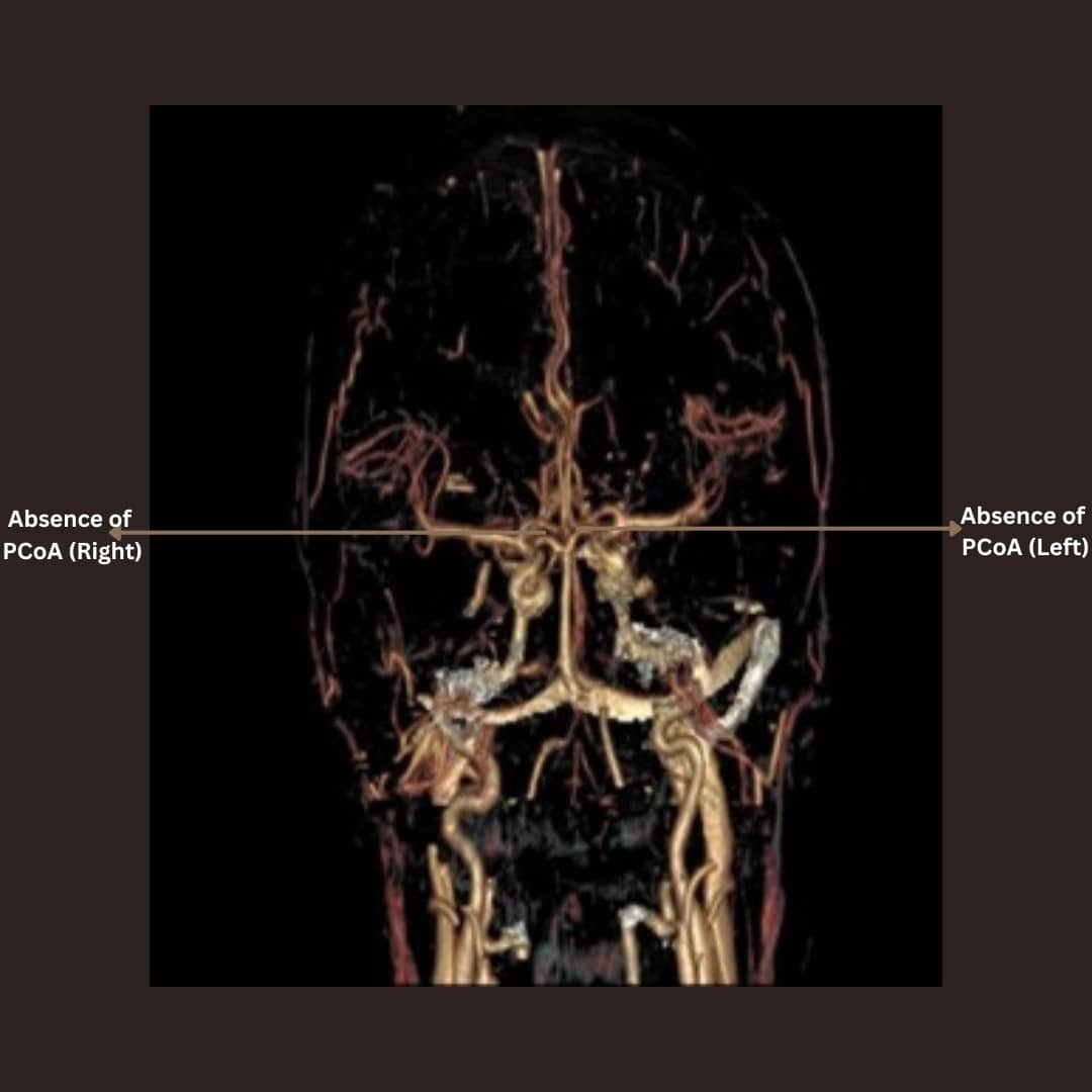
Results: Of the total 340 images evaluated, 256 (75.29%) subjects had ischemic stroke while 84 (24.71%) subjects had no stroke and were thus, considered to be apparently normal. Structural patterns in the circle of Willis mostly observed were the absence of the anterior communicating artery (10.94%) and the bilateral absence of the posterior communicating artery (10.16%).
Conclusion: Morphological patterns of the cerebral arterial circle of Willis observed, were implicated in subjects affected with stroke in the present study population.
Article Details

This work is licensed under a Creative Commons Attribution-NonCommercial-NoDerivatives 4.0 International License.
Personal views expressed by the contributors in their articles are not necessarily those of the Journal of Associated Medical Sciences, Faculty of Associated Medical Sciences, Chiang Mai University.
References
Wintermark M, Flanders AE, Velthuis B, Meuli R, Van Leeuwen M, Goldsher D, et al. Measuring elevated microvascular permeability and predicting haemorrhagic transformation in acute ischemic stroke using first-pass dynamic perfusion CT imaging. BMC Neuro. 2014; 14 (37): 6-8. doi: 10.3174/ajnr. A0539.
Navita A, Molly MP, Madhumita M. Diameter of anterior cerebral artery on MRI angiograms. Intl J Anat Res. 2016; 4(2): 2245-50. doi: 10.16965/ijar. 2016.189.
Van Overbeeke JJ, Hillen B, Tulleken CA. A comparative study of the circle of Willis in fetal and adult life; the configuration of the posterior bifurcation of the posterior communicating artery. J Anat. 1991; 176: 45-54. PMCID: PMC1260312.
Uston C. Dr. Thomas Willis famous eponym; the circle of Willis. Turk J Med Sci. 2004; 34: 271-4. doi: 10.1080/096470490512553.
Crossman AR. Vascular supply of the brain. In standing S. Gray’s anatomy. The anatomical basis of clinical practice (40th ed). Edinburg: Elsevier Churchill Living stone. 2008; 247-56. ISBN-13: 978-0443066849.
Snell SR. The blood supply of the brain and spinal cord. Clinical Neuroanatomy (7th Ed) New Delhi. Wolters Kluwer (India) Pvt. Ltd. 2010; 475-81. ISBN- 13: 978-0781794275
Sultana AA, Ara S, Rahman M, Afroz H, Fatema K, Nahar N. Variations in the sites of formation of basilar artery. Bangladesh J Anat. 2012; 10(2): 73-5. doi:10.3329/BJA.V10I2.17288.
Gunnal SA, Farooqui MS, Wabale RN. Anatomical variations of the circulus arteriosus in cadaveric human brains. J Neuro Res Intl. 2014; 68: 72-87. doi: 10.1155/2014/687281.
Singh V. Blood supply of the brain. In Textbook of clinical neuroanatomy. (2nd Ed) Elsevier Health Sciences, 2014; 172. ISBN: 8131223078, 978813122 3079.
Krabbe-Hartkamp MJ, Vander GJ. Investigation of the circle of Willis using MR angiography. Medicamundi. 2000; 44: 1. doi: 10.1148/radiology.207.1.9530305.
Kapoor K, Singh B, Dewan LI. Variations in the configuration of the circle of Willis. Anat Sci Intl. 2008; 83: 98-106. doi: 10.1111/j.1447-073X.2007.00216.x.
Nayak BS, Guru A, Devadasa SS, Rao SS. Hypoplastic plexiform right anterior cerebral artery and absence of anterior communicating artery : A case report. Forensic Med & Anat Res. 2013; 1(3): 47-9. doi: 10.4236/fmar.2013.13009.
Igiri AO, Paulinus SO, Egbe NO, Ani CC. Classical pattern of the cerebral arterial circle of Willis in a Nigerian population using contrast enhanced computed tomography scan. Intl J Sci & Engr Res. 2017; 8(8): 1204-7. Corpus ID: 235078491.
Paulinus SO, Igiri AO, Egbe NO, Ani CC, Udo-Affah GU. Evaluation of anatomical variants of the circle of Willis in a Nigerian population using Contrast Enhanced Computed Tomography (CECT) scan. Intl J Sci & Engr Res. 2017; 8(8): 2129-35.
Paulinus SO, Udoh BE, Efanga SA, Udo-Affah GU, Eru EM, Ani CC, et al. Anatomic imaging study of luminal diameter of the circle of Willis in patients with ischemic stroke. Cal J Hlth Sci. 2021; 5: 75-80. doi: 10.25259/CJHS_50_2020.
Perlmutter D, Rhoton AL. Microsurgical anatomy of the anterior cerebral–anterior communicating recurrent artery complex. J Neurosurg. 1976; 45: 259-72. doi: 10.3171/jns.1976.45.3.0259.
Rhoton AL, Saeki N, Perlmutter D, Zeal A. Microsurgical anatomy of common aneurysm sites. J Clin Neurosurg. 1979; 26: 248-306. doi: 10.1093/neurosurgery/26. cn_suppl_1.248.
Osborn AG. Diagnostic cerebral angiography. (2nd Ed). Philadelphia, Pa: Lippincort Williams & Wilkins. 1999. ISBN: 0397584040 , 9780397584048.
Luitse MJ, Van Seeters T, Horsch AD, Kool HA, Velthuis BK, Kappelle LJ, et al. Admission hyperglycaemia and cerebral perfusion deficits in acute ischemic stroke. Cereb Dis. 2013; 35: 163-7. doi: 10.1159/000346588.
Grzegorz M, Renata P, Malgorzata L. Variants of the cerebral arteries-anterior circulation. Pol J Rad. 2013; 78(3): 42-7. doi: 10.12659/PJR.889403.
Saikia B, Akash H, Pranjal P, Donboklang L, Amitav S. Circle of Willis: variant form of their embryology using gross dissection and magnetic resonance angiography. Intl J Anat & Res. 2014; 2(2): 344-53. Corpus ID: 32755817.
Bell-Gam HI, Onwuchekwa A, Iyagba AM. Improving stroke management through specialized stroke units in Nigeria: A situational review. The Nig Hlth J. 2012; 12 (2): 31-4.
Kalaria RN, Rufus A, Musafi I. Stroke injury, cognitive impairment and vascular dementia. Biochimica et Biophysica Acta. 2016; 1862(2): 915-25. doi: 10.1016/ j.bbadis.2016.01.015.
Komolafe MA, Ogunlade O, Komolafe EO. Stroke mortality in a teaching hospital in Southwest Nigeria. Trop Docs. 2007; 3: 186-8. doi: 10.1258/004947 507781524557.
Wahab KW, Okubadejo NU, Ojini FI, Danesi MA. Predictors of short-term intra-hospital case fatality following first-ever acute ischemic stroke in Nigerians. J Col Physic, Surg & Pak. 2008; 18: 755-8. PMID: 19032888.
Mukherjee D, Patil CG. Epidemiology and the global burden of stroke: Stroke and the Neurosurgeon PeerReview Report. World Neurosurg. 2011; 76(6): 85-90. doi: 10.1016/j.wneu.2011.07.023.
Desalu OO, Wahab KW, Fawale B, Olarenwaju TO, Busari OA, Adekoya AO, et al. A review of stroke admissions at a tertiary hospital in rural Southwestern Nigeria. Ann Afr Med. 2011; 10: 80-1. doi: 10.4103/ 1596-3519.82061.
Liu L, Wang D, Wong KS, Wang Y. Stroke and stroke care in China: huge burden, significant workload and a national priority. Stroke. 2011; 42(12): 3651-4. doi: 10.1161/STROKEAHA.111.635755.
Akinyemi RO, Allan L, Owolabi MO, Akinyemi JO, Ogbole G, Ajani A, et al. Profile and determinants of vascular cognitive impairment in African stroke survivors: The CogFAST Nigerian study. J Neuro Sci. 2014; 46: 241-9. doi: 10.1016/j.jns.2014.08.042.
Serefnur O. Epidemiology and the global burden of stroke-situation in Turkey. World Neurosurg. 2014; 81(6): 35-6. doi: 10.1016/j.wneu.2012.10.074.
Feigin Vl, Roth Ga, Naghavi M. Global burden of stroke and risk factors in 188 countries, during 1990- 2013: A systematic analysis for the global burden of disease study 2013. Lanc Neuro. 2016; 15(9): 913-24. doi: 10.1016/S1474-4422(16)30073-4.
World Health Organization. NCD Country Profiles. Retrieved from https://www.who.int/nmh/countries/ nga_en.pdf Accessed on the 15th June, 2018.
Camargo ECS, Furie Kl, Singhal AB, Roccatagliata L, Cunnane ME, Halpern EF, et al. Acute brain infarct: Detection and delineation with CT Angiographic source images versus non enhanced CT Scans. Rad. 2007; 244(2): 541-8. doi.org/10.1148/radiol. 2442061028.
Tan JC, Dillon WP, Liu S, Adler F, Smith WS, Wintermark M. Systematic comparison of perfusion-CT and CTangiography in acute stroke patients. Ann Neuro. 2007; 61: 533-43. doi: 10.1002/ana.21130.
Coutts SB, Hill MD, Campos CR, Choi YB, Subramaniam S, Kosior JC, et al. Recurrent events in transient ischemic attack and minor stroke. Stroke. 2008; 39: 2461-6. doi: 10.1161/STROKEAHA.107.513234.
Puetz V, Sylaja PN, Coutts SB, Hill MD, Dzialowski I, Mueller P, et al. Extent of hypo-attenuation on CT angiography source images predicts functional outcome in patients with basilar artery occlusion. Stroke. 2008; 39: 2485-90. doi: 10.1161/STROKEAHA.107.511162.
Flint AC, Kamel H, Navi BB, Rao VA, Faigeles BS, Conell C, et al. Inpatient statin use predicts improved ischemic stroke discharge disposition. Neur. 2012; 78, 21. doi: 10.1212/WNL.0b013e3182575142.
Go AS, Mozaffarian D, Roger VL. Heart disease and stroke statistics-2014 update: a report from the American Heart Association. Circulation. 2014; 129(3): 228-92. doi: 10.1161/01.cir.0000441139.02102.80.
Kramer SP. On the function of the circle of Willis. J Expl Med. 1912; 15: 348. doi: 10.1084/jem.15.4.348.
Vilmas A, Barkauskas E, Vilionskies A, Rudzinskaite J, Moikurate R. Vertebral artery hypoplasia; Importance for stroke development, the role of posterior communicating artery possibility for surgical and conservative treatment. Actamedica Lituanica. 2003; 10(2): 110-4.
Eric SS, Christopher PK, Joanna LM, Samuel SB, Micheal MM, Eric JH, et al. Radiographic absence of the posterior communicating arteries and the prediction of cognitive dysfunction after carotid endarterectomy. J Neurosurg. 2014;121(3): 593-8. doi: 10.3171/2014.5.JNS131736.
Feigin VL, Forouzanfar MH, Krishnamurthi R, Mensah GA, Connor M, Bennett DA. Global and regional burden of stroke during 1990-2010: findings from the Global Burden of Disease Study. Lanc. 2014; 383: 245-54. doi: 10.1016/s0140-6736(13)61953-4.
Thrift AG, Cadilhac DA, Thayabaranathan T. Global stroke statistics. Intl J on Stroke. 2014; 9(1): 6-18. doi: 10.1177/17474930221123175.
Obiako OR, Oparah SK, Ogunniyi A. Prognosis and outcome of acute stroke in the University College Hospital Ibadan, Nigeria. Nig J Clin Prac. 2011; 14(3): 359-62. doi: 10.4103/1119-3077.86784.
Bowen A, Wenman R, Mickelborough J, Tallis R. Dual task effects of talking while walking on velocity and balance following stroke. Ag Agg. 2001; 30: 319-23. doi: 10.1093/ageing/30.4.319.
