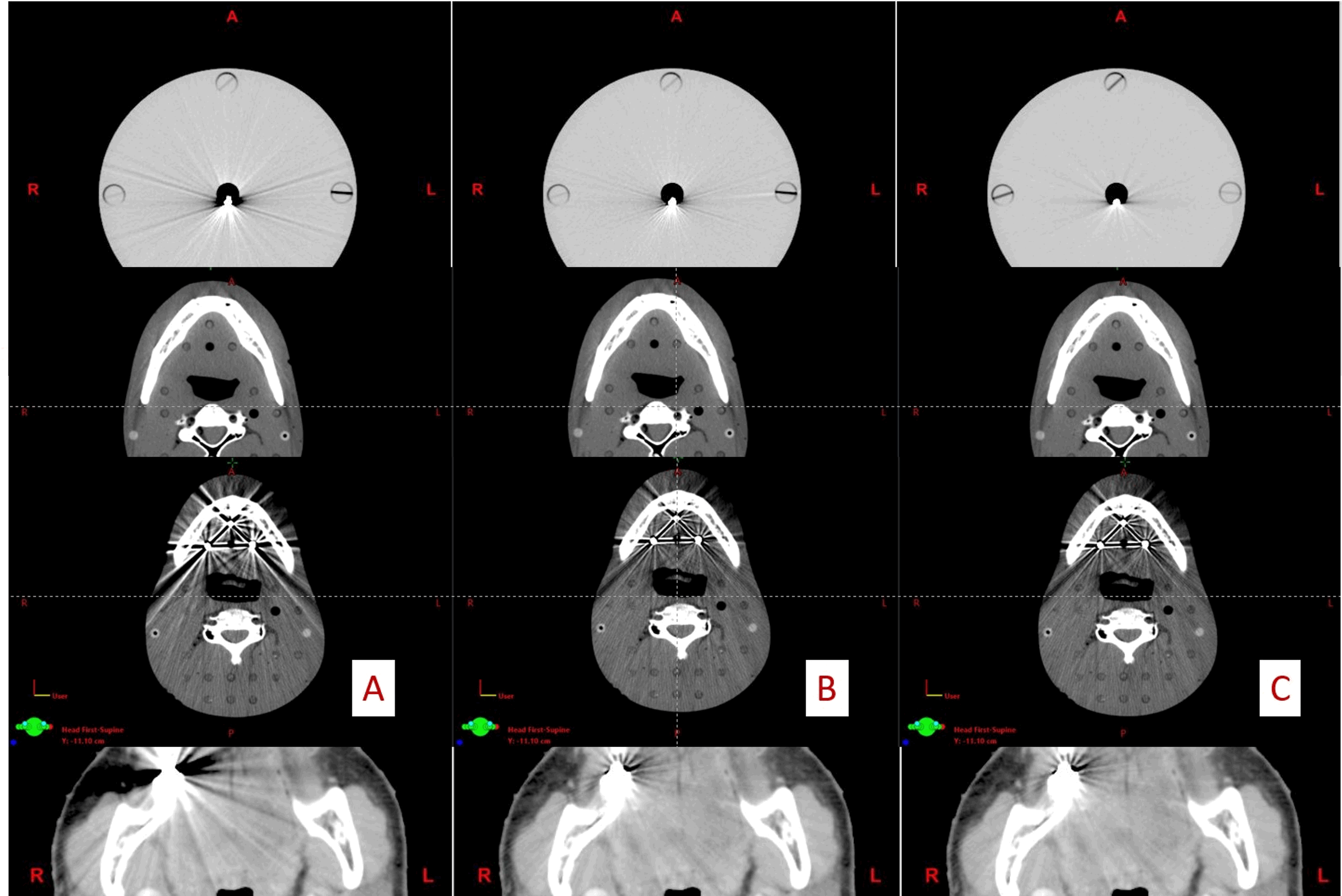
Evaluation of a new method for metal artifact reducing in computed tomographic images
Main Article Content
Abstract
Background: The common streak artifacts in computed tomographic (CT) images result from metal implants in patients. Metal artifact suppresses and obstructs diagnosis or misdiagnoses as it occurred in ten percent of the patients’ tomographic images.
Objectives: To develop the method for metal artifact reduction in CT images using MATLAB software and implement it in phantoms with the metal artifact as well as in patients with the metal artifact in the head and neck region.
Materials and methods: The new method of metal artifact reduction in CT images using MATLAB software. The homogeneous polymethylmethacrylate (PMMA) phantom, the Alderson Rando phantom, and patients with a metal implant in the head and neck region were scanned by the Philips Brilliance Big Bore CT system. Commercial orthopedic metal artifact reduction (OMAR) software and a new method software were applied to the CT images of phantoms and patients. The quantitative analysis of image quality on a metal artifact of the head and neck region was evaluated in the percent noise. The qualitative analysis in clinical imaging was evaluated in scoring by two radiologists with the same experience.
Results: In the Alderson Rando phantom, the new algorithm indicated higher efficiency in metal artifact reduction than OMAR software. In contrast, for the patient at head and neck CT images with metal artifact reduction, OMAR, and the new method showed comparable results. The new method suppressed the artifact in homogeneous PMMA, Alderson Rando phantoms, and patients with a metal implant in the head and neck region with approximately 40%, 40%, and 60% percentage of noise reduction, respectively. The qualitative analysis by two radiologists showed comparable results of OMAR and the new method.
Conclusion: The efficiency of metal artifact reduction of the new method is better than no correction and OMAR in homogeneous PMMA phantom and Alderson Rando phantom. However, the efficiency of OMAR is better than the new method, and no correction regarding the percent noise.
Article Details

This work is licensed under a Creative Commons Attribution-NonCommercial-NoDerivatives 4.0 International License.
Personal views expressed by the contributors in their articles are not necessarily those of the Journal of Associated Medical Sciences, Faculty of Associated Medical Sciences, Chiang Mai University.
References
Bushberg JT. The essential physics of medical imaging:Lippincott Williams & Wilkins; 2002.
Barrett JF, Keat N. Artifacts in CT: recognition andavoidance. Radiographics. 2004; 24(6): 1679-91. doi: 10.1148/rg.246045065
Boas FE, Fleischmann D. Evaluation of two iterativetechniques for reducing metal artifacts in computedtomography. Radiology. 2011; 259(3): 894-902. doi.org/10.1148/radiol.11101782
De Man B, Nuyts J, Dupont P, Marchal G, SuetensP, editors. Metal streak artifacts in X-ray computedtomography: a simulation study. Nuclear ScienceSymposium, 1998 Conference Record 1998 IEEE; 1998: IEEE. doi: 10.1109/23.775600
De Man B, Nuyts J, Dupont P, Marchal G, Suetens P.Reduction of metal streak artifacts in x-ray computedtomography using a transmission maximum aposteriori algorithm.IEEE Trans Nucl Sci.2000; 47(3): 977-81. doi: 10.1109/23.856534
Lemmens C, Faul D, Nuyts J. Suppression of metalartifacts in CT using a reconstruction procedure thatcombines MAP and projection completion. IEEEtransactions on medical imaging. 2009; 28(2): 250-60. doi: 10.1109/TMI.2008.929103
Popilock R, Sandrasagaren K, Harris L, Kaser KA. CTartifact recognition for the nuclear technologist. JNucl Med Technol. 2008; 36(2): 79-81. doi: 10.2967/jnmt.107.047431
Roeske JC, Lund C, Pelizzari CA, Pan X, Mundt AJ.Reduction of computed tomography metal artifactsdue to the Fletcher-Suit applicator in gynecologypatients receiving intracavitary brachytherapy.Brachytherapy. 2003; 2(4): 207-14. doi: 10.1016/j.brachy.2003.08.001
Wagenaar D, van der Graaf ER, van der Schaaf A, GreuterMJ. Quantitative comparison of commercial andnon-commercial metal artifact reduction techniquesin computed tomography. PLoS One. 2015; 10(6): e0127932. doi.org/10.1371/journal.pone.0127932
Healthcare P. Metal artifact reduction for orthopedicimplants (O-MAR). White Paper, Jan. 2012.
Mouton A, Megherbi N, Van Slambrouck K, Nuyts J,Breckon TP. An experimental survey of metal artefactreduction in computed tomography. J Xray Sci Technol.2013; 21(2): 193-226. doi: 10.3233/XST-130372
Gjesteby L, De Man B, Jin Y, Paganetti H, Verburg J,Giantsoudi D, et al. Metal artifact reduction in CT:where are we after four decades? IEEE Access. 2016; 4: 5826-49. doi: 10.1109/ACCESS.2016.2608621
Wellenberg R, Hakvoort E, Slump C, Boomsma M, MaasM, Streekstra G. Metal artifact reduction techniquesin musculoskeletal CT-imaging.Eur J Radiol. 2018; 107: 60-9. doi: 10.1016/j.ejrad.2018.08.010


