
Reliability of transversus abdominis and internal oblique muscle thickness measurement using B-mode ultrasound imaging in standing and single leg standing positions
Article Sidebar
Main Article Content
Abstract
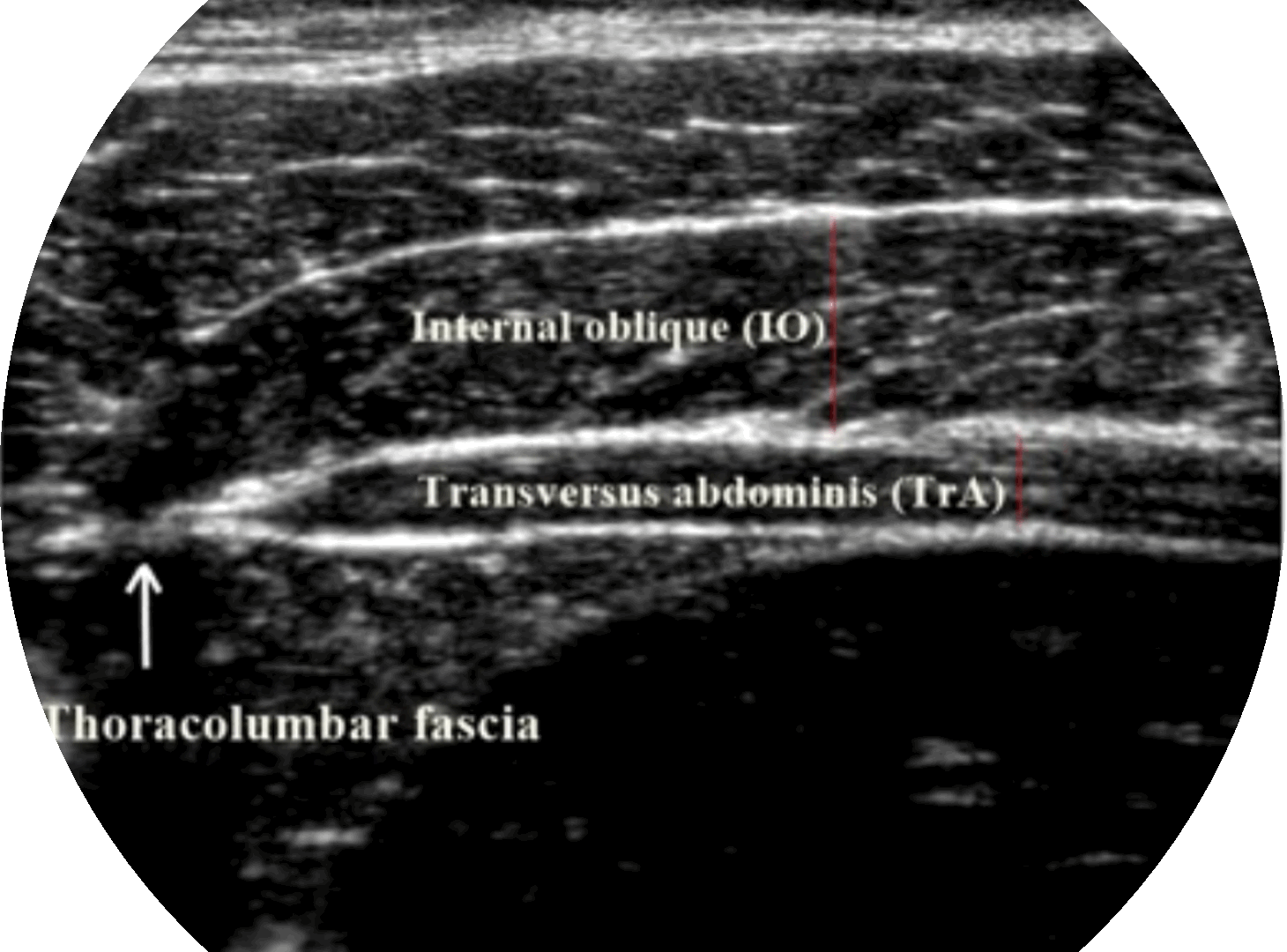
Background: The thickness of the transversus abdominis (TrA) and internal oblique (IO) muscles are measured with high reliability by musculoskeletal ultrasound imaging (MSUSI) in the supine hook-lying and walking position. However, the reliability of the procedure to measure the TrA and IO muscle thickness measurement in the functional positions such as standing and single leg standing positions have not been well established.
Objectives: To examine the reliability of MSUSI for the TrA and IO muscles thickness measurement during standing and single leg standing positions in healthy participants.
Materials and methods: Ten healthy participants (aged 24.10±2.47 years) were recruited to perform MSUSI measurement of TrA and IO muscle thickness during the resting and abdominal drawing in maneuver (ADIM) in the standing and single leg standing positions. The intra-tester reliability of MSUSI measurement was performed by the same investigator in 2 different times (24 to 72 hours apart), while the inter-tester reliability was performed by 2 investigators within the same day. The intraclass correlation coefficient (ICC) whereby ICC3,3 was used to determine the intra-tester and ICC 2,3 was used to determine the inter-tester reliability of TrA and IO muscles thickness measurement using MSUSI. The standard error of measurement (SEM) and coefficient of variation (CV) were calculated.
Results: The MSUSI measurements for TrA and IO muscles thickness showed high reliability in both standing and single leg standing positions (ICC3,3 and ICC2,3 >0.9). Moreover, the values of SEM ranged between 0.055 to 0.662 mm, MDC ranged between 0.152 to 1.834 mm and CV ranged between 0.543 to 14.001%.
Conclusion: The MSUSI measurement of TrA and IO muscles thickness could be performed with high intra and inter-tester reliability in standing and single leg standing positions among healthy individuals. The study findings on the MSUSI procedure to measure muscle thickness of TrA and IO would be useful for practice guidance in various research and clinical settings.
Article Details

This work is licensed under a Creative Commons Attribution-NonCommercial-NoDerivatives 4.0 International License.
Personal views expressed by the contributors in their articles are not necessarily those of the Journal of Associated Medical Sciences, Faculty of Associated Medical Sciences, Chiang Mai University.
References
Miura T, Yamanaka M, Ukishiro K, Tohyama H, Saito H, Samukawa M, et al. Individuals with chronic low back pain do not modulate the level of transversus abdominis muscle contraction across different postures. Man Ther. 2014; 19(6): 534-40. doi: 10.1016/j.math.2014.05.010.
Mangum LC, Henderson K, Murray KP, Saliba SA. Ultrasound assessment of the transverse abdominis during functional movement. J Ultrasound Med. 2018; 37(5): 1225-31. doi: 10.1002/jum.14466.
Mannion AF, Pulkovski N, Toma V, Sprott H. Abdominal muscle size and symmetry at rest and during abdominal hollowing exercises in healthy control subjects. J Anat. 2008; 213(2): 173-82. doi: 10.1111/ j.1469-7580.2008.00946.x.
Rankin G, Stokes M, Newham DJ. Abdominal muscle size and symmetry in normal subjects. Muscle Nerve. 2006; 34(3): 320-6. doi: 10.1002/mus.20589.
Sitilertpisan P, Joseph L, Paungmali A, Paungmali U, Chunchai T. Investigation of the contraction ratio of transversus abdominis and internal oblique muscles during lumbopelvic stability test. MLTJ. 2020; 10: 86. doi: 10.32098/mltj.01.2020.10.
Oliva-Lozano JM, Muyor JM. Core muscle activity during physical fitness exercises: a systematic review. Int J Environ Res Public Health. 2020; 17(12). doi: 10.3390/ijerph17124306.
Watson T, Graning J, McPherson S, Carter E, Edwards J, Melcher I, et al. Dance, balance and core muscle performance measures are improved following a 9-week core stabilization training program among competitive collegiate dancers. Int J Sports Phys Ther. 2017; 12(1): 25-41.
Gnat R, Saulicz E, Miądowicz B. Reliability of real-time ultrasound measurement of transversus abdominis thickness in healthy trained subjects. Eur Spine J. 2012; 21(8): 1508-15. doi: 10.1007/s00586-012-2184-4.
Hides JA, Miokovic T, Belavý DL, Stanton WR, Richardson CA. Ultrasound imaging assessment of abdominal muscle function during drawing-in of the abdominal wall: an intrarater reliability study. J Orthop Sports Phys Ther. 2007; 37(8): 480-6. doi: 10.2519/jospt.2007.2416.
Koppenhaver SL, Hebert JJ, Fritz JM, Parent EC, Teyhen DS, Magel JS. Reliability of rehabilitative ultrasound imaging of the transversus abdominis and lumbar multifidus muscles. Arch Phys Med Rehabil. 2009; 90(1): 87-94. doi: 10.1016/j.apmr.2008.06.022.
Koppenhaver SL, Hebert JJ, Parent EC, Fritz JM. Rehabilitative ultrasound imaging is a valid measure of trunk muscle size and activation during most isometric sub-maximal contractions: a systematic review. Aust J Physiother. 2009; 55(3): 153-69. doi: 10.1016/s0004-9514(09)70076-5.
Mangum LC, Sutherlin MA, Saliba SA, Hart JM. Reliability of ultrasound imaging measures of transverse abdominis and lumbar multifidus in various positions. Pm R. 2016; 8(4): 340-7. doi: 10.1016/j.pmrj.2015.09.015.
Lee DH, Hong SK, Lee YS, Kim CH, Hwang JM, Lee Z, et al. Is abdominal hollowing exercise using real-time ultrasound imaging feedback helpful for selective strengthening of the transversus abdominis muscle?: A prospective, randomized, parallel-group, comparative study. Medicine (Baltimore). 2018; 97(27). doi: 10.1097/md.0000000000011369.
Walter SD, Eliasziw M, Donner A. Sample size and optimal designs for reliability studies. Stat Med. 1998; 17(1): 101-10. doi: 10.1002/(sici)1097-0258(19980115)17:1<101::aid-sim727>3.0.co;2-e.
van Melick N, Meddeler BM, Hoogeboom TJ, Nijhuis-van der Sanden MWG, van Cingel REH. How to determine leg dominance: The agreement between self-reported and observed performance in healthy adults. PLoS One. 2017; 12(12): e0189876. doi: 10.1371/journal.pone.0189876.
Teyhen DS, Miltenberger CE, Deiters HM, Del Toro YM, Pulliam JN, Childs JD, et al. The use of ultrasound imaging of the abdominal drawing-in maneuver in subjects with low back pain. J Orthop Sports Phys Ther. 2005; 35(6): 346-55. doi: 10.2519/ jospt.2005.35.6.346.
Springer BA, Mielcarek BJ, Nesfield TK, Teyhen DS. Relationships among lateral abdominal muscles, gender, body mass index, and hand dominance. J Orthop Sports Phys Ther. 2006; 36(5): 289-97. doi: 10.2519/jospt.2006.2217.
Prentice CLS, Milanese S, Massy-Westropp N, Maranna S. The reliability of rehabilitative ultrasound to measure lateral abdominal muscle thickness: A systematic review and meta-analysis. Musculoskelet Sci Pract. 2021; 53: 102357. doi: 10.1016/j.msksp.2021.102357.
