
Role of computed tomographic enterography for evaluation of small bowel diseases: A cross-sectional study
Article Sidebar
Main Article Content
Abstract
Background: Computed tomographic enterography (CTE) is a newer non-invasive modality having distinct advantages over conventional CT and capsule endoscopy.
Objectives: This technique allows faster evaluation of small bowel diseases in the endoscopically inaccessible segments. Being an operator-independent procedure, CTE is widely available and allows a better depiction of extra enteric complications. The aim is to evaluate CTE features of various small bowel diseases and the role of 2% mannitol for adequate small bowel distension.
Materials and methods: A cross-sectional study comprising 105 patients had presented with small bowel diseases. Patients in the age group of 10 to 85 years with complaints of fever, abdominal pain, nausea, vomiting, altered bowel habits, loss of appetite and loss of weight were included in this study. CTE images were analyzed to compare the diagnosis with the available histopathological and ultrasonography results.
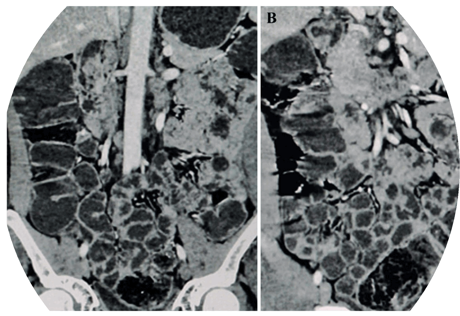
Results: Among the study population, the majority had presented CTE features such as symmetrical wall thickening (53.3%), peri-bowel inflammatory changes (61%), mucosal hyperenhancement (39%), and mural stratification, i.e., target sign (33.3%). The majority of diagnoses of CTE were ileocecal tuberculosis (11.5%), small bowel inflammation (7.6%), and Crohn’s disease (6.7%). Other conditions such as small bowel neoplastic masses, diverticula, ischemic bowel disease, bowel strictures, intussusception, and ulcerative colitis.
Conclusion: CTE has the vital role of first-line modality in the work-up of suspected small intestinal diseases and helps evaluate disease activity before endoscopy, particularly in inaccessible segments. It allows a better depiction of extra enteric complications of the bowel.
Article Details

This work is licensed under a Creative Commons Attribution-NonCommercial-NoDerivatives 4.0 International License.
Personal views expressed by the contributors in their articles are not necessarily those of the Journal of Associated Medical Sciences, Faculty of Associated Medical Sciences, Chiang Mai University.
References
Mazzeo S, Caramella D, Battolla L, Melai L, Masolino P, Bertoni M, et al. Crohn disease of the small bowel: Spiral CT evaluation after oral hyperhydration with isotonic solution. J Comput Assist Tomogr
[Internet]. 2001; 25: 612-6. DOI: 10.1097/00004728200107000-00017.
Tochetto S, Yaghmai V. CT Enterography: Concept, Technique, and Interpretation. Radiol Clin N Am. 2009; 47: 117-32. DOI: 10.1016/j.rcl.2008.10.007.
Megibow AJ, Babb JS, Hecht EM, Jennie J, Cho, BS, Carmela HMS, et al. Evaluation of bowel distention and bowel wall appearance by using neutral oral contrast agent for multi-detector row CT. Radiology. 2006; 238: 87-95. DOI: 10.1148/radiol.2381041985.
Saibeni S, Rondonotti E, Iozzelli A, et al. Maurizio Vecchi. Imaging of the small bowel in Crohn’s disease: A review of old and new techniques. World J Gastroenterol. 2007; 13: 3279-87. DOI: 10.3748/wjg.v13.i24.3279.
Macari M, Megibow AJ, Balthazar EJ. A pattern approach to the abnormal small bowel: observations at MDCT and CT enterography. Am J Roentgenol. 2007; 188: 1344-55. DOI: 10.2214/AJR.06.0712.
Wold PB, Fletcher JG, Johnson CD, Sandborn WJ. Assessment of Small Bowel Crohn Disease: Noninvasive Peroral CT Enterography Compared with Other Imaging Methods and Endoscopy-Feasibility Study. Radiology. 2003; 229: 275-81. DOI: 10.1148/radiol.2291020877.
Boudiaf M, Jaff A, Soyer P, Bouhnik Y, Hamzi L, Rymer R. Small-Bowel Diseases: Prospective Evaluation of Multi–Detector Row Helical CT Enteroclysis in 107 Consecutive Patients. Radiology. 2004; 233: 338-44.
Lim BK, Bux SI, Rahmat K, Lam SY, Liew YW. Evaluation of bowel distension and mural visualisation using neutral oral contrast agents for multidetector-row computed tomography. Singap Med J. 2012; 53(11): 732-6.
Zhang LH, Zhang SZ, Hu HJ, Min Gao, Ming Zhang, Qian Cao, et al. Multi-detector CT enterography with iso-osmotic mannitol as oral contrast for detecting small bowel disease. World J Gastroenterol. 2005; 11(15): 2324-9. DOI: 10.3748/wjg.v11.i15.2324.
Masselli G, Gualdi G. CT and MR enterography in evaluating small bowel diseases: when to use which modality? Abdom Imaging. 2013; 38: 249-59.
Misra RN, Bajaj SK. Role of CT enterography in evaluation of small bowel disorders. Int J of Res Med Sci. 2019; 7: 537-543. DOI: https://dx.doi.org/10.18203/2320-6012.ijrms20185504.
Goldberg HI, Gore RM, Margulis AR, Moss AA, Baker EL. Computed tomography in the evaluation of Crohn disease. Am J Roentgenol. 1983; 140: 277-82. DOI: 10.2214/ajr.140.2.277.
Balthazar EJ. CT of the gastrointestinal tract: principles and interpretation. Am J Roentgenol. 1991; 156: 23-32. DOI: 10.2214/ajr.156.1.1898566.
Horton KM, Fishman EK. The current status of multidetector row CT and three- dimensional imaging of the small bowel. Radiol Clin N Am. 2003; 41: 199-212. DOI: 10.1016/s0033-8389(02)00121-5.
Horton KM, Eng J, Fishman EK. Normal enhancement of the small bowel: evaluation with spiral CT. J Comput Assist Tomogr. 2000; 24: 67-71. doi: 10.1097/00004728-200001000-00014.
Dos-Santos CHM, Menezes JNS, Nunes TF, Martins LA. CT enterography in the evaluation of Crohn’s disease. J Coloproctol (Rio J). 2015; 35(4): 217-22. DOI: 10.1016/j.jcol.2015.06.006
