
Audit of computed tomography examinations from two selected radio-diagnostic centers in South-South Nigeria
Article Sidebar
Main Article Content
Abstract
Background: Despite the fact that the number of CT exams is small among all radiography investigations, a high amount of medical radiation exposure comes from CT application. Most developed nations have adopted regular audits to ensure optimization of ionizing radiations in the CT examinations, but on the contrary, it has infrequently been performed in developing countries like Nigeria.
Objectives: This study was designed to carry out an audit of CT examinations at two selected diagnostic centers in the South-South region of Nigeria.
Materials and methods: This study was a retrospective cross-sectional study conducted in two radiological facilities, which involved 210 tomographs of the chest, head, and abdomen, selected using a convenient method. The CT examinations were done using the departmental protocols and the generated data were analyzed statistically using descriptive statistics.
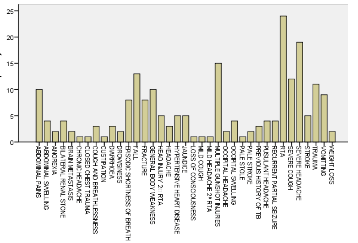
Results: Head examination was the most commonly performed CT examination (56.7%), followed by abdominal 28.6 % and the least 14.8% was chest. The most common indication was a road traffic accident (RTA) 11.4%. The distribution of the type of CT machine that was used for the study showed that the Toshiba machine was used for most of the subjects 132 (62.9%) followed by Optima CT660 78 (37.1%). It was seen that 48.6% of the study used 0.75s, 40.5% used 0.5s, 10.5% used 0.35s and only 0.5% used 1s scan time. The effective doses were adult head (2.31±0.14), chest (4.65±0.21), abdominal (7.70±0.17), pediatric head (2.81±0.21), and pediatric chest (9.96±0.12).
Conclusion: The carrying out of clinical audits is imperative to ensure both safeties of patients and diagnostic accuracy.
Article Details

This work is licensed under a Creative Commons Attribution-NonCommercial-NoDerivatives 4.0 International License.
Personal views expressed by the contributors in their articles are not necessarily those of the Journal of Associated Medical Sciences, Faculty of Associated Medical Sciences, Chiang Mai University.
References
Mettler F, Bhargavan M, Faulkner K., Giller D, Gray J, Ibbott G. Frequency, Radiation dose and comparison with other radiation sources. Radiol Society North Am (RSNA). 2009; 259: 520-31.
Aydin P, Gokce K, Tolga I, Emine B, Figen B, Tolga O, Sadi G. Patient doses from CT examination in Turkey. Diagn Interventional Radiol J. 2015; 21: 428-34.
Elliot A. (2009). Issues in Medical exposure. J Radiol. 2009; 29: 107-21.
Chiaghanam NO, Nzotta CC, Enweani IB (2019) Radiation Risk Assessment of Soil in Idomi, Cross River State, Nigeria. Asian J Appl Sci. 2019; 7(1): 27-35.
Brenner D, Hall E. Computed Tomography-An increasing source of radiation exposure. American J Radiol. 2007; 357(2): 2277-84.
Chanda R, Bwanga O, Sindaza N, Mercy NC, Chisha M (2020) Audit of completion of Computed Tomography (CT) request forms at the Cancer disease hospital (CDH) of Zambia. Med J Zambia. 2020; 47(4): 289-96.
Chiaghanam NO, Nwoyi IE. Paediatric entrance surface doses in routine X-ray examinations in three radiology facilities within South-South, Nigeria. Sci Technol. 2020; 6: 12-9.
Chiaghanam NO, Esien-umo EO, Egbe NO, Asuquo CF, Akpanama D.P (2022a) Efficacy of shielding structure in Computed Tomography Suite in selected radiodiagnostic centres in Uyo metropolis Akwa Ibom state Nigeria. Inter J Sci and Engineering Res. 2022b; 13(3): 215-33.
Chiaghanam NO, Esien-umo E, Ogolodom MP, Asuquo C, Maurice C, Omita E, Ugwuanyi DC, Ezugwu EE. Safety of Ionizing Radiation in selected Conventional X-ray Diagnostic centres in Calabar and Uyo metropolises, Nigeria. Euro Sci J. 2022b; 18(21): 1-9.
Ogbole GI, Obed R. Ra diation dose in computed Tomography: Need for optimization and application of dose reference levels in Nigeria. West Afri J Radiol. 2004; 21(1):1-16.
Liang CR, Chen PXH, Kapor J, Ong MKL, Quek ST, Kapor SC. Establishment of Institutional diagnostic reference level for CT with automated tracking software. J Med Radiat Sci. 2017; (64): 82-9.
Small R., Surujpaul P.P, Chakraborty S. (2019) Patient Dose Audit in Computed Tomography at Cancer Institute of Guyana. J Med Diagn Meth. 2019; 18(1): 1-13.
Newman B, GangulyA, Kim JE, Robinson T. Comparison of different methods of calculating CT radiation effective dose in children. AJR Am J Roentgenol. 2012: 199: W232-W9.
European Union. European Commission guidelines on clinical audit from medical radiological practice. Publication 159. 2004.
International Atomic Energy Agency. Optimization of the radiological protection of patients undergoing radiography, fluoroscopy and computed tomography. Annals of IAEA coordinated research project. Austria. 2004.
International Atomic Energy Agency. Dosimetry in Diagnostic Radiology for pediatric patients. Human Health Series-IAEA no. 24. 2013.
Bongartz G, Golding S, Jurik A, Leonardi, M. Van Persijn. Quality criteria for multislcie computed tomography. Results from a European concerted action on CT (FIGM-CT-2000-20078) Appendix B. European Field Survey on MSCT. 2004.
DDM2. Study on European population doses from medical exposure. 2012.
Friberg E, Borretzen I, Olerud M. Dual and Multidetector row CT׃ what about the doses?” presented at the Radiation protection symposium of the Northwest European Radiation Protection societies, Utrecht, the Netherlands. 2003.
Foly, S., McEntee, M., Rainford L. Establishment of CT diagnostic reference levels in Ireland. British J Radiol. 2012; 85: 1390-7.
Prokop M, Galanski M. Spiral and multislice computed tomography of the body. Thieme, New York, NY. 2003; p. 133-60.
Hidajat N, Wolf M, Nunnemann A, Liersch P, Gebauer B. Survey of conventional and spiral CT doses. J Radiol. 2001; 218: 395-40.
Shrimpton P, Hillier M, Lewes M, Dunn M. National Radiological Protection Board (NRPB): doses from Computed Tomography examinations in the UK-2003 review. Document NRPB-W67. Chilton England. 2005.
Shrimpton, P., Hillier, M., Lewis, M., Dunn, M. National survey of doses from CT in the UK: 2003. British J Radiol. 2006; 79: 768-980.
Papadimitrious D, Perris A, Maneton A, Molfetas M, Panagiotakis N. A survey on 14 computed tomography scanners in Greece and 32 scanners in Italy. Examination frequencies, dose reference values, effective doses and doses to organs. J Radiat Protect Dosimetry. 2003; 104: 47-53.
Tsapaki V, Aldrich J, Sharma R, Staniszewska M, Krisanachinda A. Dose reduction in CT while maintaining diagnostic confidence: Diagnostic dose reference levels at routine head, chest and abdominal CT- IAEA- coordinated research project. 2006; 240: 828-34.
Klang E., Beytelman A., Greenberg D., Or J., Zimlichman E. Overuse of Head CT examinations for the investigation of minor head truma: Analysis contributing factor. J Am Coll Radiol. 2017; 14(2): 171-6.
Ploussi A, Efstathopoulos P E. Importance of establishing protection culture in radiology department. World J Radiol. 2016; 8: 142-7.
