
Hepatic lesion detectability in abdomen computed tomography: Investigation in low kVp single energy and low keV virtual monochromatic images generated from dual energy computed tomography using task-based image quality assessment in phantom
Article Sidebar
Main Article Content
Abstract
Background: Low tube potential single-energy (SE) and virtual monochromatic image (VMI) dual energy (DE) abdomen CT images both improve the hepatic lesion detection by increasing the liver lesion contrast on images.
Objectives: To study the performance of low kVp single energy and low keV virtual monochromatic images (VMI) generated from dual energy acquisition to detect the hepatic lesion on abdominal CT imaging in phantom.
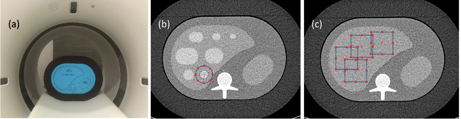
Materials and methods: The anthropomorphic liver with nodule inserted phantom with extension rings simulating the small, medium, and large patient was scanned under the SECT acquisition by varying the kVp from 70-120 kVp and for DECT acquisitions, three kVp combinations (80/-,90/-, and 100/Sn150-kVp). The series of 40-,50-,60-, and 70-keV VMI were generated from DECT data set. All images were used to assess the task-based image quality; task transfer function (TTF), noise power spectrum (NPS), and detectability index (d’) with the diagnostic task to detect 15 mm diameter hyperattenuating hepatic lesion.
Results: The result showed that the TTF was higher in low kVp SECT while the lesion contrast was higher at low keV VMI -DECT. The noise magnitude remained constant for all kVp values in SECT, but it was dramatically increased as decreased the energy level from 70- to 40-keV in VMI-DECT. The fav of NPS shifted to higher frequency when increasing the kVp and when increasing the energy level of VMI. The obtained d’ was highest in low kVp SECT at 70-or 80-kVp.
Conclusion: The low kVp SECT provided the highest d’ than that in low keV VM image from DECT in all phantom sizes where the highest d’ was found at 70 kVp-SECT for small and medium phantom and at 80 kVp for large phantom. For SECT, reduced the kVp to 70- or 80-kVp improved the detectability index. The kVp combination in DECT impacts to the d’ of VMI; at small phantom, the highest d’ for each keV VMI was found at 80/Sn150 kVp acquisition and at larger phantom size, higher kVp on tube “A” is more favored.
Article Details

This work is licensed under a Creative Commons Attribution-NonCommercial-NoDerivatives 4.0 International License.
Personal views expressed by the contributors in their articles are not necessarily those of the Journal of Associated Medical Sciences, Faculty of Associated Medical Sciences, Chiang Mai University.
References
Sung H, F erlay J, Sie gel RL , Laversanne M, Soerjomataram I, Jemal A, et al. Global Cancer Statistics 2020: GLOBOCAN Estimates of Incidence and Mortality Worldwide for 36 Cancers in 185 Countries. CA Cancer J Clin. 2021; 71(3): 209-49.
Choi JY, Lee JM, Sirlin CB. CT and MR imaging diagnosis and staging of hepatocellular carcinoma: part II. Extracellular agents, hepatobiliary agents, and ancillary imaging features. Radiology. 2014; 273(1): 30-50.
Marrero JA, Kulik LM, Sirlin CB, Zhu AX, Finn RS, Abecassis MM, et al. Diagnosis, Staging, and Management of Hepatocellular Carcinoma: 2018 Practice Guidance by the American Association for the Study of Liver Diseases. Hepatology. 2018; 68(2): 723-50.
Kim DW, Choi SH, Kim SY, Byun JH, Lee SS, Park SH, et al. Diagnostic performance of MRI for HCC according to contrast agent type: a systematic review and meta-analysis. Hepatol Int. 2020; 14(6): 1009-22.
Ehman EC, Guimarães LS, Fidler JL, Takahashi N, Ramirez-Giraldo JC, Yu L, et al. Noise reduction to decrease radiation dose and improve conspicuity of hepatic lesions at contrast-enhanced 80-kV hepatic CT using projection space denoising. Am J Roentgenol. 2012;198(2): 405-11.
Lv P, Liu J, Chai Y, Yan X, Gao J, Dong J. Automatic spectral imaging protocol selection and iterative reconstruction in abdominal CT with reduced contrast agent dose: initial experience. Eur Radiol. 2017; 27(1): 374-83.
Hanson GJ, Michalak GJ, Childs R, McCollough B, Kurup AN, Hough DM, et al. Low kV versus dual-energy virtual monoenergetic CT imaging for proven liver lesions: what are the advantages and trade-offs in conspicuity and image quality? A pilot study. Abdom Radiol (NY). 2018;43(6): 1404-12.
Lee YJ, Lee JM, Lee JS, Lee HY, Park BH, Kim YH, et al. Hepatocellular carcinoma: diagnostic performance of multidetector CT and MR imaging-a systematic review and meta-analysis. Radiology. 2015;275(1):97-109.
Samei E, Bakalyar D, Boedeker KL, Brady S, Fan J, Leng S, et al. Performance evaluation of computed tomography systems: summary of AAPM task group 233. Med Phys. 2019; 46(11): e735-e56.
Chen B, Christianson O, Wilson JM, Samei E. Assessment of volumetric noise and resolution performance for linear and nonlinear CT reconstruction methods. Med Phys. 2014; 41(7): 071909.
Verdun F, Racine D, Ott J, Tapiovaara M, Toroi P, Bochud F, et al. Image quality in CT: From physical measurements to model observers. Physica Medica. 2015; 31(8): 823-43.
Shuman WP, Green DE, Busey JM, Mitsumori LM, Choi E, Koprowicz KM, et al. Dual-energy liver CT: effect of monochromatic imaging on lesion detection, conspicuity, and contrast-to-noise ratio of hypervascular lesions on late arterial phase. Am J Roentgenol. 2014; 203(3): 601-6.
Choi JY, Lee JM, Sirlin CB. CT and MR imaging diagnosis and staging of hepatocellular carcinoma: part I. Development, growth, and spread: key pathologic and imaging aspects. Radiology. 2014; 272(3): 635-54.
Thun M, Linet MS, Cerhan JR, Haiman CA, Schottenfeld D. Cancer epidemiology and prevention: Oxford University Press; 2017.
Solomon J, Wilson J, Samei E. Characteristic image quality of a third generation dual-source MDCT scanner: Noise, resolution, and detectability. Med Phys. 2015; 42(8): 4941-53.
Michalak G, Grimes J, Fletcher J, Halaweish A, Yu L, Leng S, et al. Selection of optimal tube potential settings for dual-energy CT virtual mono-energetic imaging of iodine in the abdomen. Abdom Radiol (NY). 2017; 42(9): 2289-96.
Greffier J, Si-Mohamed S, Dabli D, de Forges H, Hamard A, Douek P, et al. Performance of four dual-energy CT platforms for abdominal imaging: a task-based image quality assessment based on phantom data. Eur Radiol. 2021; 31(7): 5324-34.
Greffier J, Frandon J, Sadate A, Akessoul P, Belaouni A, Beregi JP, et al. Impact of four kVp combinations available in a dual‐source CT on the spectral performance of abdominal imaging: A task‐based image quality assessment on phantom data. J Appl Clin Med Phys. 2021; 22(8): 243-54.
Cester D, Eberhard M, Alkadhi H, Euler A. Virtual monoenergetic images from dual-energy CT: systematic assessment of task-based image quality performance. Quant Imaging Med Surg. 2022; 12(1): 726.
Solomon J, Wilson J, Samei E. Characteristic image quality of a third generation dual‐source MDCT scanner: Noise, resolution, and detectability. Med Phys. 2015; 42(8): 4941-53.
