
ภาวะก้อนแคลเซียมจากยูรีเมียในผู้ป่วยฟอกเลือดเรื้อรัง: การรักษาด้วยโซเดียมไธโอซัลเฟต
Article Sidebar
Main Article Content
บทคัดย่อ
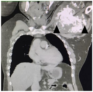
ภาวะก้อนแคลเซียมจากยูรีเมียเป็นภาวะแทรกซ้อนที่พบได้ไม่บ่อยในผู้ป่วยที่ได้รับการบบำบัดทดแทนไต โดยทั่วไปภาวะนี้จะสัมพันธ์กับระดับแคลเซียมและฟอสเฟตที่สูงในเลือดส่งผลให้เกิดก้อนแคลเซียมสะสมอยู่ที่บริเวณข้อต่างๆ ในร่างกาย สามารถพบภาวะนี้ได้บ่อยขึ้นในผู้ป่วยที่ได้รับการฟอกเลือดหรือล้างไตมาเป็นระยะเวลานาน ซึ่งภาวะก้อนแคลเซียมจากยูรีเมียนี้จะส่งผลกระทบต่อระบบกล้ามเนื้อและกระดูก ทำให้มีอาการปวดเมื่อย คุณภาพชีวิตแย่ลง และผู้ป่วยบางคนอาจไม่สามารถทำงานได้ การทบทวนวรรณกรรมในเชิงลึกแสดงให้เห็นว่าผู้ป่วยส่วนใหญ่ไม่ตอบสนองต่อการรักษาด้วยการควบคุมอาหาร การรับประทานยา หรือการปรับวิธีการฟอกเลือด บทความนี้รายงานผู้ป่วยชายอายุ 29 ปี ที่ได้รับการฟอกเลือดด้วยเครื่องไตเทียมมาเป็นระยะเวลาหลายปี มีก้อนหลายก้อนที่ใต้ผิวหนังบริเวณไหล่ซ้าย มีอาการปวด และขนาดของก้อนใหญ่ขึ้นเรื่อย ๆ ตลอดระยะเวลา 3 ปีที่ผ่านมา ในช่วงแรกผู้ป่วยได้รับการวินิจฉัยว่าเป็นก้อนจากกรดยูริกที่พบในโรคเก๊าท์ จึงได้รับการรักษาด้วยยาลดกรดยูริกขนาดสูงอยู่ระยะหนึ่ง แต่ขนาดของก้อนไม่ลดลงแต่กลับโตขึ้น ผู้ป่วยได้รับการตรวจเพิ่มเติมซึ่งรวมไปถึงการส่งตรวจทางพยาธิวิทยาของชิ้นเนื้อจากก้อน จนในที่สุดได้รับการวินิจฉัยว่าเป็นภาวะก้อนแคลเซียมจากยูรีเมีย ผู้ป่วยได้รับการรักษาด้วยการใช้น้ำยาฟอกเลือดที่มีความเข้มข้นของแคลเซียมต่ำ รับประทานยาจับฟอสเฟตที่ไม่มีแคลเซียมเป็นส่วนประกอบ และยาโซเดียมไธโอซัลเฟตทางหลอดเลือดดำ หลังจากติดตามผลการรักษาเป็นระยะเวลา 6 เดือน พบว่าอาการปวดทุเลาลง ขนาดของก้อนเล็กกว่าร้อยละ 50 และคุณภาพชีวิตโดยรวมของผู้ป่วยดีขึ้นอย่างชัดเจน
Article Details

อนุญาตภายใต้เงื่อนไข Creative Commons Attribution-NonCommercial-NoDerivatives 4.0 International License.
บทความนี้ตีพิมพ์ภายไต้การอนุญาต CC BY-NC-ND 4.0 ซึ่งอนุญาตให้สามารถใช้บทความนี้พื่อวัตถุประสงค์ใดๆ ก็ตามที่ไม่ใช่เชิงพาณิชย์ โดยต้องมีการอ้างถึงที่มาของบทความอย่างครบถ้วน ใครก็ตามสามารถคัดลอกและแจกจ่ายทุกส่วนของบทความนี้โดยไม่ต้องขออนุญาตจากผู้ประพันธ์หรือสมาคมโรคไตแห่งประเทศไทย
เอกสารอ้างอิง
Floege J. When man turns to stone: Extraosseous calcification in uremic patients. Kidney Int. 2004;65:2447-62.
Raja DL, Podymow T, Barre T. Tumoral calcinosis in a peritoneal dialysis patient. Kidney Int. 2006;70(11):1887.
Kai MC, Cheuk CS, Angela YM. Uremic tumoral calcinosis. Nephrol Dial Transplant. 2004;19:505-6.
Chakarun CJ, Talkin B, White EA, Miriam R, Philip W. Tumoral calcinosis: sonographic sedimentation sign. J Clin Ultrasound. 2011;39:367-70.
Hamada J, Tamai K, Ono W, Saotome K. Uremic tumoral calcinosis in hemodialysis patients: clinicopathological findings and identification of calcific deposits. J Rheumatol. 2006;33:119–26.
Cofan F, Garcia S, Combalia A, Campistol JM, Oppenheimer F, Ramon R. Uremic tumoral calcinosis inpatients receiving long-term hemodialysis therapy. J Rheumatol. 1999;26:379–85.
Ibels LS. The pathogenesis of metastatic calcification in uremia. Prog Biochem Pharmacol. 1980;17:242–50
Ibrahim M, Kabbaja A, Issouani M, Hassanai A. Tumoral calcinosis: Diffuse multifocal form in hemodialysis patients. Two case reports. Orthop Traumatol Surg Res. 2017;103(5): 815–20.
Guermazi A, Grigoryan M, Cordoliani F, Kerob D. Unusually diffuse idiopathic calcinosis cutis. Clin Rheumatol. 2007;26: 268–70.
Rafaelsen S, Johansson S, Raeder H, Bjerknes R. Long-term clinical outcome and phenotypic variability in hyperphosphatemic familial tumoral calcinosis and hyperphosphatemic hyperostosis syndrome caused by a novel GALNT3 mutation; case report and review of the literature. BMC Genet. 2014;15:98.
Malberti F, Ravani P. The choice of the dialysate calcium concentration in the management of patients on hemodialysis and hemodiafiltration. Nephrol Dial Transplant. 2003;7:37–40.
LeGeros RZ, Contiguglia SR, Alfrey AC. Pathological calcifications associated with uremia: two types of calcium phosphate deposits. Calcif Tissue Res. 1973;13(3);173-85.
Van Straten A, Hoogeveen EK, Khan SHM, Arthur M. Unusual presentation of tumoral calcinosis in chronic renal failure: a case report. Eur J Radiol. 2005;53(2):81-5.
Drueke TB, Touam M, Thorney Brown D, Rostand SG. Extraskeletal calcification in patients with chronic kidney failure. Adv Nephrol. 2000;30:333–56.
Yaerim Kim, Eunah Hwang, Sungbae Park. Case report Resolution of uremic tumoral calcinosis in a patient on peritoneal dialysis with long-term low-calcium dialysate treatment. Kidney Res Clin Pract. 2014;33:226–28.
Aoun A, Baubion E, Banydeen R, Djiconkpode l, Ekindi N, Urena Torres, et al. Incidence and characteristics of calciphylaxis in Martinique (2006–2012). Ann Dermatol Venereol. 2014;141(12):743–9.
Hug I, Gunçaga J. Tumoral calcinosis with sedimentation sign. Br J Radiol. 1974;47(562): 734-6.
Chakarun CJ, Talkin B, White EA, Romero M, Ralls PW. Tumoral calcinosis: sonographic sedimentation sign. J Clin Ultrasound. 2011;39(6):367-70.
Zhen H, Kyung JS, Dillon C, Wing C, Jim S. Imaging features of soft-tissue calcifications and related diseases: A Systematic approach. Korean J Radiol. 2018;19(6):1147-60.
Shen X, Chen W, Wang X, Chen J. Uremic tumoral calcinosis in a patient on hemodialysis. Intern Med. 2012;51(11):1443.
Papadakis JT, Patrikarea A. Sodium thiosulfate in the treatment of tumoral calcifications in a hemodialysis patient without hyperparathyroidism. Nephron 1996; 72:308-12.
Fathi I, Sakr M. Review of tumoral calcinosis: a rare clinicopathological entity. World J Clin Cases. 2014;2:409–14.
